universitÀ degli studi di padova -...
TRANSCRIPT
UNIVERSITÀ DEGLI STUDI DI PADOVA
Sede Amministrativa: Università degli Studi di Padova
Dipartimento di Scienze Sperimentali Veterinarie
SCUOLA DI DOTTORATO DI RICERCA IN SCIENZE
VETERINARIE
INDIRIZZO SCIENZE BIOMEDICHE VETERINARIE E
COMPARATE
CICLO XXII
TESI DI DOTTORATO DI RICERCA:
BIOACTIVE PEPTIDES FROM MILK PROTEINS: FOCUSING ON PEPTIDES DISPLAYING
IMMUNOMODULATORY ACTIVITY
Direttore della Scuola: Ch. mo Prof. Massimo Morgante
Supervisore: Ch. mo Prof. Alessandro Negro
Dottoranda: Daniela Regazzo
INDEX
I
ABBREVIATIONS LIST ........................................................................................................................ V
SOMMARIO ...................................................................................................................................... 1
SUMMARY ....................................................................................................................................... 3
1. AIM OF THE RESEARCH ........................................................................................................... 5
2. REVIEW OF LITERATURE .......................................................................................................... 7
2.1. MILK AND MILK-DERIVED PRODUCTS .................................................................................................... 7
2.2. BIOACTIVE PEPTIDES ......................................................................................................................... 9
2.2.1. Definition ............................................................................................................................ 9
2.2.2. Mechanisms of production of bioactive peptides ............................................................. 10 2.2.2.1. Bioactive peptide release during gastrointestinal digestion through the action of digestive
enzymes or microbial enzymes of the intestinal flora ........................................................................... 11 2.2.2.2. Bioactive peptide release during milk processing trough the action of microbial enzymes ..... 12 2.2.2.3. Bioactive peptide release during milk processing trough the action of a single purified enzyme
or a combination of selected enzymes .................................................................................................. 13 2.2.3. Mechanisms of action of bioactive peptides ..................................................................... 14
2.2.4. Commercial dairy products and ingredients with health or function claims based on
bioactive peptides ....................................................................................................................... 15
2.3. BIOACTIVITIES OF INTEREST .............................................................................................................. 17
2.3.1. ACE-inhibition ................................................................................................................... 17 2.3.1.1. Physiology of ACE-inhibition ..................................................................................................... 18 2.3.1.2. ACE-inhibitory peptides derived from milk ............................................................................... 20 2.3.1.3. Microorganisms and enzymes for the production of fermented milk with ACE-inhibitory
activity ................................................................................................................................................... 25 2.3.2. Immunomodulation .......................................................................................................... 30
2.3.2.1. Overview of the physiology of the immune system .................................................................. 30 2.3.2.2. Immunomodulatory peptides derived from milk ...................................................................... 36 2.3.2.3. Microorganisms for the production of fermented milk with immunomodulatory activity ...... 40 2.3.2.4. Two examples of immunomodulatory peptides derived from milk proteins ........................... 42
2.3.2.4.1. YGG peptide ...................................................................................................................... 43 2.3.2.4.2. β-CN (193-209) peptide ................................................................................................... 44
2.4. BIOACTIVE PEPTIDE DIGESTION ......................................................................................................... 44
2.4.1. Physiology of the digestion of proteins and peptides ....................................................... 45 2.4.1.1. The digestion of bioactive peptides derived from milk proteins .............................................. 51
2.4.2. Digestion Models .............................................................................................................. 52 2.4.2.1. The brush-border membrane vesicles ...................................................................................... 60
2.5. BIOACTIVE PEPTIDE ABSORPTION ....................................................................................................... 61
2.5.1. Physiology of the absorption of proteins and peptides..................................................... 61
2.5.2. Physical and chemical characteristics of potentially absorbable bioactive peptides ........ 66 2.5.2.1. The absorption of bioactive peptides derived from milk proteins ............................................ 67
2.5.3. Absorption models ............................................................................................................ 68 2.5.3.1. The Caco-2 cell line model ........................................................................................................ 73
EXPERIMENT 1: FERMENTED MILK FROM ENTEROCOCCUS FAECALIS TH563 OR LACTOBACILLUS
DELBRUECKII BULGARICUS LA2 MANIFESTS DIFFERENT DEGREES OF ACE-INHIBITORY AND
IMMUNOMODULATORY ACTIVITIES .............................................................................................. 77
3.1. INTRODUCTION ............................................................................................................................. 77
3.2. MATERIALS AND METHODS ............................................................................................................. 78
3.2.1. Chemicals and Reagents ................................................................................................... 78
3.2.2. Bacteria culture ................................................................................................................. 79 3.2.3. Separation of the peptide fraction .................................................................................... 79
3.2.4. ACE-inhibitory activity ....................................................................................................... 80
3.2.5. Bovine peripheral blood lymphocytes proliferation .......................................................... 80
3.3. RESULTS ...................................................................................................................................... 81
3.4. DISCUSSION .................................................................................................................................. 83
3.5. TAKE-HOME MESSAGE .................................................................................................................... 85

INDEX
II
EXPERIMENT 2: EFFECTS OF YGG ON (CONCANAVALIN A-INDUCED) PROLIFERATION AND IL2 AND
INFg EXPRESSION OF BOVINE PERIPHERAL BLOOD LYMPHOCYTES ................................................ 87
4.1. INTRODUCTION .............................................................................................................................. 87
4.2. MATERIALS AND METHODS .............................................................................................................. 89
4.2.1. Chemicals and Reagents .................................................................................................... 89
4.2.2. BPBL Harvesting and Propagation ..................................................................................... 89
4.2.3. Part 1: BPBL proliferation .................................................................................................. 89
4.2.4. Part 2: IL2 and INFγ gene expression ................................................................................. 90
4.2.5. Data analysis ..................................................................................................................... 92
4.3. RESULTS ....................................................................................................................................... 93
4.3.1. BPBL proliferation .............................................................................................................. 93
4.3.2. IL2 and INFγ gene expression............................................................................................. 94
4.4. DISCUSSION ................................................................................................................................... 96
4.5. TAKE-HOME MESSAGE ..................................................................................................................... 99
EXPERIMENT 3: STUDY OF THE BIOACTIVE PROPERTIES AND THE TRANSPORT OF THE PEPTIDE Β-CN
(193-209), A 17-RESIDUES PEPTIDE OF BOVINE Β-CASEIN, THROUGH CACO-2 MONOLAYERS ..... 101
5.1. INTRODUCTION ............................................................................................................................101
5.2. MATERIALS AND METHODS ............................................................................................................102
5.2.1. Chemicals and Reagents ..................................................................................................102
5.2.2. Preparation of β-CN (193-209) ........................................................................................103
5.2.3. Cell Culture .......................................................................................................................103
5.2.4. Transepithelial transport studies .....................................................................................104
5.2.5. Effects of β-CN (193-209) on cellular viability .................................................................105
5.2.6. Effects of β-CN (193-209) on tight junctions: TJ-stabilizing activity ................................106
5.2.7. RP-HPLC-ESI/MS analyses ................................................................................................106
5.2.8. Assessment of β-CN (193-209) hydrolysis ........................................................................107
5.2.9. Data analysis ...................................................................................................................108
5.3. RESULTS .....................................................................................................................................108
5.3.1. Transepithelial transport of β-CN (193-209) across the Caco-2 cells ...............................108
5.3.2. Influence of Gly-Pro, Cytochalasin D and wortmannin on β-CN (193-209) transport ......111
5.3.3. Influence of β-CN (193-209) on Caco-2 TJ stability and permeability ..............................112
5.3.4. Influence of β-CN (193-209) on Caco-2 viability ..............................................................113
5.4. DISCUSSION .................................................................................................................................114
5.5. TAKE-HOME MESSAGE ...................................................................................................................119
EXPERIMENT 4: ASSESSMENT OF DIGESTION OF THE PEPTIDE Β-CN (193-209), A 17-RESIDUES
PEPTIDE OF BOVINE Β-CASEIN, ON BRUSH BORDER MEMBRANE VESICLES .................................. 121
6.1. INTRODUCTION ............................................................................................................................121
6.2. MATERIALS AND METHODS ............................................................................................................122
6.2.1. Chemicals and Reagents ..................................................................................................122
6.2.2. Preparation of β-CN (193-209) ........................................................................................122
6.2.3. Preparation of BBMV .......................................................................................................122
6.2.4. Assessment of Β-CN (193-209) digestion by pBBMV and wpBBMV.................................124
6.2.5. Identification of peptides by RP-HPLC-ESI/MS .................................................................125
6.2.6. Data analysis ...................................................................................................................125
6.3. RESULTS .....................................................................................................................................126
6.3.1. Assessment of digestion ..................................................................................................126
6.3.2. Kinetics of digestion .........................................................................................................127
6.3.3. Identification of peptides generated during digestion ....................................................130
6.4. DISCUSSION .................................................................................................................................132
6.5. TAKE-HOME MESSAGE ...................................................................................................................134
GENERAL DISCUSSION .................................................................................................................. 137
7.1. STUDIES ON THE DIGESTION AND ABSORPTION OF BIOACTIVE PEPTIDES ....................................................137
7.2. THE EVALUATION OF THE IMMUNOMODULATORY ACTIVITY OF BIOACTIVE PEPTIDES ....................................139

INDEX
III
7.3. FUTURE PERSPECTIVES ON THE PRODUCTION OF DAIRY FOOD WITH ACE-INHIBITORY AND IMMUNOMODULATORY
PROPERTIES ....................................................................................................................................... 141
CONCLUSIONS .............................................................................................................................. 143
ACKNOWLEDGEMENTS ................................................................................................................ 147
WEB REFERENCES ......................................................................................................................... 149
REFERENCES ................................................................................................................................. 150

ABBREVIATIONS LIST
V
ABBREVIATIONS LIST ACE Angiotensin converting enzyme
A. oryzae Aspergillus oryzae
AUC Area Under the Curve
B. lactis Bifidobacterium lactis
BALT Bronchus-Associated Lymphoid Tissue
BBMV Brush border membrane vesicles
BPBL Bovine peripheral blood lymphocytes
C. cardunculus Cynara cardunculus
α-CN, β-CN, κ-CN α-casein, β-casein, κ-casein
conA Concanavalin A
E. faecalis Enterococcus faecalis
DBP Diastolic blood pressure
DMEM Dulbecco’s modified Eagle medium
DMSO Dimethyl sulfoxide
EDTA Ethylenediaminetetraacetic acid
FAAs Free Amino Acids
FCS Fetal Calf Serum
FOSHU Food Specified Health Use
g Gravity acceleration (9.8 m/s2)
GALT Gut-Associated Lymphoid Tissue
GI Gastrointestinal
L-Glu L-Glutamine
HA Hippuric Acid
HBSS Hank’s Buffered Salt Solution
HEPES Hydroxyethyl Piperazine Ethane Sulphonic Acid
ABBREVIATIONS LIST
VI
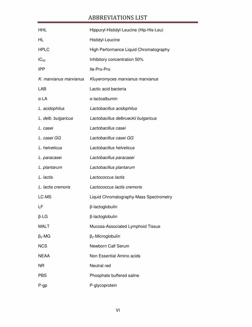
HHL Hippuryl-Histidyl-Leucine (Hip-His-Leu)
HL Histidyl-Leucine
HPLC High Performance Liquid Chromatography
IC50 Inhibitory concentration 50%
IPP Ile-Pro-Pro
K. marxianus marxianus Kluyeromyces marxianus marxianus
LAB Lactic acid bacteria
α-LA α-lactoalbumin
L. acidophilus Lactobacillus acidophilus
L. delb. bulgaricus Lactobacillus delbrueckii bulgaricus
L. casei Lactobacillus casei
L. casei GG Lactobacillus casei GG
L. helveticus Lactobacillus helveticus
L. paracasei Lactobacillus paracasei
L. plantarum Lactobacillus plantarum
L. lactis Lactococcus lactis
L. lactis cremoris Lactococcus lactis cremoris
LC-MS Liquid Chromatography-Mass Spectrometry
LF β-lactoglobulin
β-LG β-lactoglobulin
MALT Mucosa-Associated Lymphoid Tissue
β2-MG β2-Microglobulin
NCS Newborn Calf Serum
NEAA Non Essential Amino acids
NR Neutral red
PBS Phosphate buffered saline
P-gp P-glycoprotein
ABBREVIATIONS LIST
VII
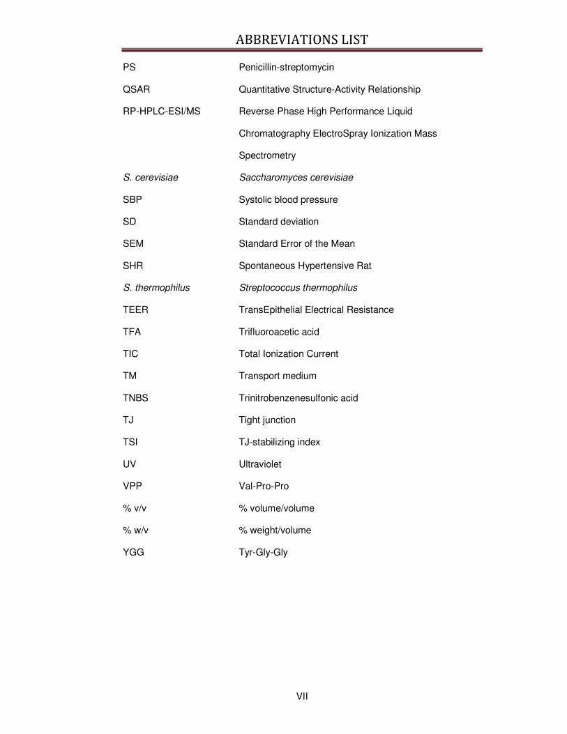
PS Penicillin-streptomycin
QSAR Quantitative Structure-Activity Relationship
RP-HPLC-ESI/MS Reverse Phase High Performance Liquid
Chromatography ElectroSpray Ionization Mass
Spectrometry
S. cerevisiae Saccharomyces cerevisiae
SBP Systolic blood pressure
SD Standard deviation
SEM Standard Error of the Mean
SHR Spontaneous Hypertensive Rat
S. thermophilus Streptococcus thermophilus
TEER TransEpithelial Electrical Resistance
TFA Trifluoroacetic acid
TIC Total Ionization Current
TM Transport medium
TNBS Trinitrobenzenesulfonic acid
TJ Tight junction
TSI TJ-stabilizing index
UV Ultraviolet
VPP Val-Pro-Pro
% v/v % volume/volume
% w/v % weight/volume
YGG Tyr-Gly-Gly
SOMMARIO
1
SOMMARIO
I peptidi bioattivi derivati dal latte costituiscono una parte importante del latte, in
grado di influenzare lo stato di salute. Attualmente nel latte e nei suoi derivati sono
stati identificati e caratterizzati peptidi ad azione oppioide, anti-trombotica, anti-
ipertensiva, immunomodulatoria, antiossidante, antimicrobica, anticancro, stimolanti
l’assorbimento di minerali e la crescita. In questa tesi particolare attenzione è stata
rivolta ai peptidi bioattivi ad attività ACE-inibitoria e immunomodulatoria.
Nell’Esperimento 1 Enterococcus faecalis TH563 (E. faecalis TH563) e
Lactobacillus delbrueckii subsp. bulgaricus LA2 (L. delb. bulgaricus LA2), due ceppi
batterici isolati da formaggi tradizionali del Nord Italia, sono stati caratterizzati per la
loro capacità di produrre latti fermentati arricchiti in attività ACE-inibitoria e
immunomodulatoria. I risultati preliminari hanno dimostrato che il ceppo E. faecalis
TH563 è in grado di produrre un latte fermentato con elevata attività ACE-inibitoria
mentre il ceppo L. delb. bulgaricus LA2 produce un latte fermentato con attività
immunomodulatoria su linfociti bovini.
Per meglio comprendere i meccanismi che regolano l’attività immunomodulatoria
manifestata dal latte fermentato, nell’Esperimento 2 sono stati riportati i risultati di un
esperimento atto a valutare gli effetti immunomodulatori del peptide bioattivo YGG.
Tale tripeptide può essere generato durante il processo di fermentazione del latte
dalla proteina α–lattoalbumina mediante l’azione proteolitica degli enzimi batterici, e
quindi anche durante la fermentazione operata dai ceppi E. faecalis TH563 e L.
delb. bulgaricus LA2. YGG è stato somministrato a linfociti isolati da sangue bovino
e ne è stata studiata la capacità di modulare la proliferazione dei linfociti e
l’espressione (RNA) di due citochine (IL2 e INFγ) in diverse condizioni di coltura
(presenza/assenza di attivatori della proliferazione, diverse concentrazioni di siero
bovino). Lo studio ha dimostrato che il peptide YGG è in grado di modulare la

SOMMARIO
2
proliferazione delle cellule e che tale modulazione è influenzata dalle condizioni di
coltura ma non sembra essere mediata dalle citochine oggetto di studio.
Un fattore importante che limita l’impiego su larga scala di alimenti con proprietà
bioattive è la biodisponibilità dei peptidi portatori di tali bioattività. I fattori che
maggiormente influenzano la biodisponibilità dei peptidi sono la resistenza alla
digestione operata dagli enzimi gastrointestinali e la possibilità che tali peptidi
possano essere assorbiti dall’epitelio intestinale. A questo scopo, negli Esperimenti
3 e 4 sono stati esaminati il profilo di digestione e i meccanismi di assorbimento del
peptide β-CN (193-209). β-CN (193-209) è un peptide bioattivo lungo e idrofobico,
derivato dalla β-caseina ed è già stato isolato e identificato in diversi prodotti derivati
dal latte come yogurt e latte fermentati. Tale peptide possiede inoltre diverse attività
immunomodulatorie. Il profilo di digestione di tale peptide e i meccanismi di
assorbimento intestinale sono stati studiati in modelli in vitro adatti a rappresentare
la mucosa intestinale, come le vescicole della membrana a orletto a spazzola
(BBMV) e la linea cellulare Caco-2. Tali esperimenti hanno dimostrato che il peptide
viene assorbito intatto dalle cellule Caco-2, probabilmente attraverso un trasporto
mediato da vescicole.
In conclusione, il contributo principale di questa tesi di dottorato è stato il fornire
nuova conoscenza sui prodotti derivati dal latte ad azione bioattiva. Più
specificatamente, questa tesi ha permesso di ottenere nuove informazioni sui
meccanismi di produzione dei peptidi bioattivi derivati dal latte, sul loro meccanismo
d’azione e sulla loro stabilità nel sistema gastrointestinale. Infine, i risultati ottenuti
hanno contribuito a generare nuove idee che potranno costituire nuovi spunti per
futuri progetti di ricerca.
SUMMARY
3
SUMMARY
Milk-derived peptides are milk components able to influence specific physiological
functions, finally acting on body health condition. At present, the bioactivities
described for milk-derived peptides include opiate, antithrombotic, antihypertensive,
immunomodulating, antioxidative, antimicrobial, anticancer, mineral-carrying and
growth-promoting activities. In this thesis, special attention has been given to
bioactive peptides with ACE-inhibitory and immunomodulatory activities.
In the Experiment 1 Enterococcus faecalis TH563 (E. faecalis TH563) and
Lactobacillus delbrueckii subsp. bulgaricus LA2 (L. delb. bulgaricus LA2), two
bacterial strains isolated from traditional North Eastern Italy dairy products, have
been evaluated for their ability to produce fermented milk rich in ACE-inhibitory and
immunomodulatory activities. The preliminary results obtained from this experiment
demonstrated that E. faecalis TH563 produced a fermented milk with high ACE-
inhibitory activity while L. delb. bulgaricus LA2 showed an immunomodulatory
activity on bovine lymphocytes.
To better understand the mechanisms underlying the immunomodulatory activity of
fermented milks, in the Experiment 2 the immunomodulatory effects of the milk-
derived bioactive tri-peptide YGG have been examined. YGG could be generated
during milk fermentation from α–lactalbumin hydrolysis operated by bacterial
enzymes, so it could be present in milk fermented by L. delb. bulgaricus LA2. YGG
has been administered to purified peripheral blood lymphocytes in different culture
conditions (presence/absence of activators of lymphocyte proliferation, different
concentration of newborn calf serum) and its effects on lymphocyte proliferation and
cytokine RNA expression (IL2 and INFγ) have been analyzed. YGG modulated
lymphocytes proliferation, in a manner dependent from culture conditions but its
effects did not seem mediated by the modulation of IL2 or INFγ RNA expression.
SUMMARY
4
An important limiting factor of the large-scale diffusion of food carrying potential
bioactivities is the bioavailability of the peptides responsible of such bioactivities.
The main factors influencing the bioavailability of peptides are the resistance to
digestion enzymes of and the absorption by the intestinal epithelium. In the
Experiments 3 and 4 the sensitivity to gastrointestinal enzymes and the mechanisms
of absorption of the peptide β-CN (193-209) have been evaluated. β-CN (193-209)
is a long hydrophobic peptide derived from β-casein that has been already isolated
and identified from fermented milks and yogurt and displayed immunomodulatory
properties. The pattern of digestion and the mechanisms of absorption have been
evaluated in well-known in vitro models for the intestinal epithelium, as the brush
border membrane vesicles (BBMV) and the Caco-2 cell line. The results of these
studies demonstrated that the β-CN (193-209) peptide is absorbed intact by the
Caco-2 monolayer, probably via a vesicles-mediated mechanism.
In conclusion, the main contribution of this PhD thesis was to provide new
knowledge about milk-derived products with bioactivities. In particular, original
contributions are in relation to the mechanisms by which milk-derived bioactive
peptides are generated, express their bioactivities, and their fate in the
gastrointestinal tract. As a result, new questions have arisen on this area that could
constitute the objective of further research programs in the future.
AIM OF THE RESEARCH
5
1. AIM OF THE RESEARCH
The present thesis aimed to elucidate the function and the bioavailability of bioactive
peptides present in milk or in milk-derived products, with the purpose to identify the
crucial aspects that have to be taken into consideration for an efficient production of
bioactive peptides from milk proteins. In this context, special attention has been
given to the immunomodulatory activity and to specific milk-derived peptides
associated to this bioactivity.
The critical aspects that have to be considered for the production of bioactive
peptides from milk proteins are various, as the mechanisms of release of bioactive
peptides from milk proteins and the bioavailability of the peptides in the body and in
this PhD thesis the more relevant have been evaluated.
First, the mechanisms of generation of the bioactivities from the raw milk, notably
the effect of the bacterial strain on the digestive phenomena intervening in the
production of fermented milks rich in ACE-inhibitory and immunomodulatory
activities, have been studied.
Secondly, the factors involved in the bioavailability of bioactive peptides, as the
resistance to digestion and the mechanism of absorption by the intestinal epithelium,
have been assessed in two well established in vitro models for the intestinal
epithelium, as brush border membrane vesicles and Caco-2 cell line.
Finally, the mechanisms of action by which the immunomodulatory peptides
manifest their activity once into the target organism have been characterized in
bovine peripheral blood lymphocytes.
REVIEW OF LITERATURE
7
2. REVIEW OF LITERATURE
2.1. Milk and milk-derived products
Milk is the secretion of the mammary gland, containing approximately 5% lactose,
3.1 protein, 4% lipid and 0.7% minerals. The components of milk provide critical
nutritive elements, immunological protection, and biologically active substances to
both neonates and adults. It is not surprising, therefore, that the nutritional value of
milk is high.
From an objective viewpoint, it seems logical that a lactating animal, as well as
providing vital early nutrition, would also protect the health of its offspring via the
biochemical influences of its milk. In particular, the notion that components within
milk can influence and direct the physiological development of the offspring, as its
environmental exposure increases, is now widely accepted [1]. The concept of
bovine milk as a biologically active fluid is therefore not new [2], but the identification
of factors within bovine milk that may be relevant to improving human health, and
the potential development of bovine milk-containing preparations into products with
proven health-promoting properties, certainly is [1].
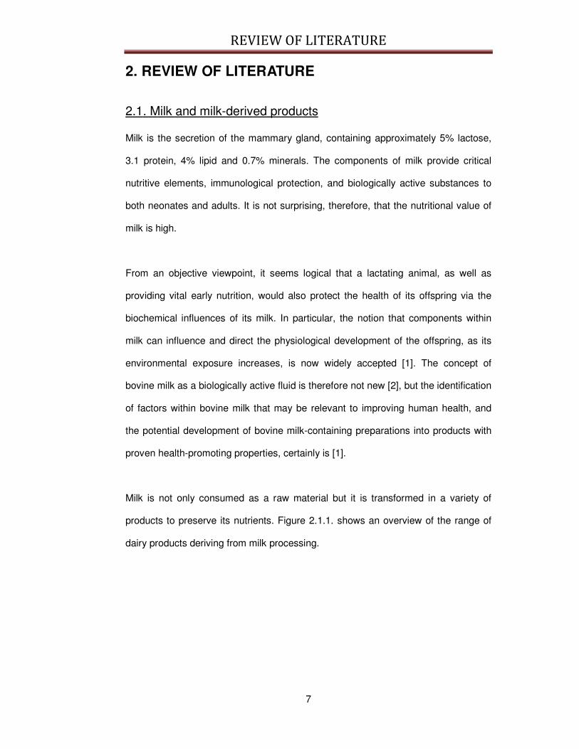
Milk is not only consumed as a raw material but it is transformed in a variety of
products to preserve its nutrients. Figure 2.1.1. shows an overview of the range of
dairy products deriving from milk processing.
REVIEW OF LITERATURE
8
Fig 2.1.1. Overview of the range of dairy products deriving from milk processing. From: http://www.foodsci.uoguelph.ca/dairyedu/home.html.
Among all the dairy products, milk fermentation and cheese making are the oldest
methods used to extend the shelf-life of milk, and they have been practiced by
human beings for thousands of years [3]. Recently, numerous scientific works [4-7]
have demonstrated and confirmed that the consumption of fermented milk and
cheeses manifests health beneficial effects that go beyond the nutritional value.
Indeed, fermented milk consumption has been associated with reduction of serum
cholesterol [8], antihypertensive [5] and osteoprotective [9] effects. The mechanisms
of action responsible of these properties have been investigated and have been
attributed to the numerous bioactive peptides contained in milk and/or released
during milk processing.

REVIEW OF LITERATURE
9
It is not surprising that in recent years intense research interest has been focused
on identifying biologically active components within bovine milk and milk-derived
products, and characterising the way by which mammalian physiological function is
modulated by these components. Not surprisingly, a significant proportion of this
research has sought to characterise the potential of bovine milk, milk products or
milk components to influence some of the most important body physiological
functions, as blood pressure [10-12], the immune system [13-15], and the resistance
to the infections [16]. For example, there is now a substantial body of evidence to
suggest that major components of bovine milk, as well as several constituents or
even yogurt and cheese, can regulate blood pressure in humans [5, 17]. The most
significant advances in this field have been made over the last five to ten years, and
this review will focus primarily on the recent advances and current knowledge in this
rapidly expanding field. Moreover, particular attention is given to the milk-derived
bioactive peptides responsible of some important health properties.
2.2. Bioactive peptides
2.2.1. Definition
Accordingly to a widely shared definition [18], a bioactive dietary substance is “a
food component that can affect biological processes or substrates and, hence, have
an impact on body function or condition and ultimately health”. In addition, dietary
substances should give a measurable biological effect in the range of doses it is
usually assumed in the food and this bioactivity should be measured at a
physiologically realistic level [9].
Following this definition, milk-derived bioactive peptides are milk components able to
influence some physiological functions, finally acting on body health condition.
Moreover, among the numerous bioactive substances studied up to now, increasing
interest is focused on milk-derived bioactive peptides because at present, bovine
REVIEW OF LITERATURE
10
milk, cheese and dairy products seem to be extremely important sources of
bioactive peptides derived from food.
2.2.2. Mechanisms of production of bioactive peptides
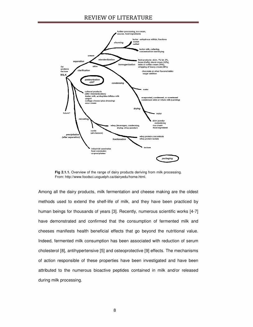
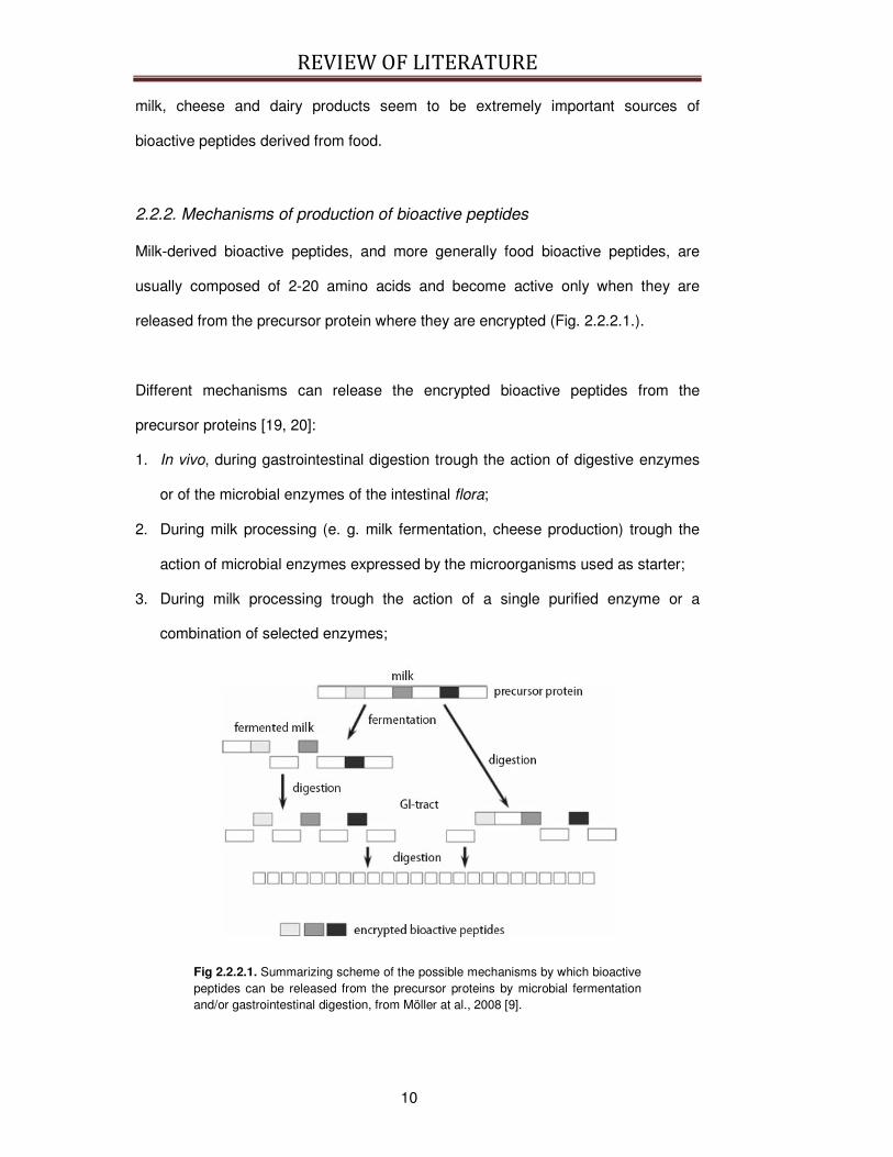
Milk-derived bioactive peptides, and more generally food bioactive peptides, are
usually composed of 2-20 amino acids and become active only when they are
released from the precursor protein where they are encrypted (Fig. 2.2.2.1.).
Different mechanisms can release the encrypted bioactive peptides from the
precursor proteins [19, 20]:
1. In vivo, during gastrointestinal digestion trough the action of digestive enzymes
or of the microbial enzymes of the intestinal flora;
2. During milk processing (e. g. milk fermentation, cheese production) trough the
action of microbial enzymes expressed by the microorganisms used as starter;
3. During milk processing trough the action of a single purified enzyme or a
combination of selected enzymes;
Fig 2.2.2.1. Summarizing scheme of the possible mechanisms by which bioactive peptides can be released from the precursor proteins by microbial fermentation and/or gastrointestinal digestion, from Möller at al., 2008 [9].
REVIEW OF LITERATURE
11
2.2.2.1. Bioactive peptide release during gastrointestinal digestion
through the action of digestive enzymes or microbial enzymes of the
intestinal flora
Bioactive peptides may be released in vivo during gastrointestinal digestion. These
bioactive peptides are mostly the result of the degradation of casein with several
proteases such as pepsin, trypsin or chymotrypsin. At present, despite some
experimental works on the stimulation of gastrointestinal digestion of eggs and meat
proteins [21, 22], the production of milk-derived bioactive peptides in vivo during
digestion remains unclear. Before dietary proteins can be cleaved by pancreatic
proteases in the intestine, they pass through the stomach, in which food can remain
for up to several hours depending on its composition and degree of particle
reduction during mastication. In the gastric juice, the proteins undergo degradation
by HCl and pepsin. While the peptide products resulting from milk proteins digestion
with site-specific pancreatic proteases, such as trypsin or chymotrypsin are well
investigated [23, 24], there are only few papers regarding this primary step of human
digestion of milk proteins [25, 26].
During gastrointestinal digestion, bioactive peptides may be released from the
precursor protein throughout the whole intestine. In fact, proteins contained in food
matrices enter the stomach through the cardiac orifice and they are further
denatured and partially degraded by the combined action of HCl and pepsin. This
first digestion step operated on proteins in the stomach permits the consequent
action of the enzymes present in the small intestine, which are the main responsible
of protein hydrolysis. Thus, bioactive peptides are predominantly released in this
portion of the gastrointestinal tract. Microbial enzymes of the resident gut flora can
act only on milk proteins that reach the large intestine intact or only partially
degraded [9]. Compared to the gastrointestinal enzymes, microbial enzymes, either
REVIEW OF LITERATURE
12
in the intestine or used as starter during milk processing, use different cleavage
sites. Thus, bioactive peptides liberated by microbial enzymes may differ from those
released by digestive enzymes. It remains to be elucidated if these bioactive
peptides released by the resident flora of the large intestine could be absorbed and
in which extent. In addition, when the bioactive peptides are released by bacterial
enzymes during milk fermentation, they could be the target of the action of
gastrointestinal enzymes and they may release other bioactive peptides [9].
Moreover, it has been demonstrated that the peptidic profile of milk proteins is
significantly different after microbial fermentation, suggesting that microbial
proteolysis can be a potential source of bioactive peptides during milk processing
[27].
2.2.2.2. Bioactive peptide release during milk processing trough the
action of microbial enzymes
Many industrially utilized dairy starter cultures are highly proteolytic. Bioactive
peptides can, thus, be generated by the starter and non-starter bacteria used in the
manufacture of fermented dairy products. The proteolytic system of lactic acid
bacteria (LAB), e.g. Lactococcus lactis, Lactobacillus helveticus and L. delb.
bulgaricus, is already well characterized. This system consists of a cell wall-bound
proteinase and a number of distinct intracellular peptidases, including
endopeptidases, aminopeptidases, tripeptidases and dipeptidases [28]. Rapid
progress has been made in recent years to elucidate the biochemical and genetic
characterization of these enzymes.
Many recent articles and book chapters have reviewed the release of various
bioactive peptides from milk proteins through microbial proteolysis [27, 29, 30]. In
addition, a number of studies have demonstrated that hydrolysis of milk proteins by
REVIEW OF LITERATURE
13
digestive and/or microbial enzymes may produce peptides with immunomodulatory
activities [31].
2.2.2.3. Bioactive peptide release during milk processing trough the
action of a single purified enzyme or a combination of selected
enzymes
The most common way to produce bioactive peptides is through enzymatic
hydrolysis of whole protein molecules. ACE-inhibitory peptides and calcium-binding
phosphopeptides, for example, are most commonly produced by trypsin [32-35].
Moreover, ACE-inhibitory peptides have recently been identified in the tryptic
hydrolysates of bovine αs2-casein [36] and in bovine, ovine and caprine k-casein
macropeptides [37]. Other digestive enzymes and different enzyme combinations of
proteinases - including alcalase, chymotrypsin, pepsin and thermolysin as well as
enzymes from bacterial and fungal sources - have also been utilized to generate
bioactive peptides from various proteins [19, 38].
Proteolytic enzymes isolated from LAB have been successfully employed to release
bioactive peptides from milk proteins. Yamamoto and colleagues [39] reported that
casein hydrolyzed by the cell wall-associated proteinase from L. helveticus CP790
showed antihypertensive activity in spontaneously hypertensive rats. Several ACE-
inhibitory peptides and one antihypertensive peptide were isolated from the
hydrolysate. Maeno et al. [40] hydrolyzed casein using the same proteinase and
identified a β-casein-derived antihypertensive peptide, the fragment β-CN (169-175),
whose amino acidic sequence is KVLPVPQ. In a recent study, Mizuno and
colleagues [41] measured the ACE-inhibitory activity of casein hydrolysates upon
treatment with nine different commercially available proteolytic enzymes. Among
these enzymes, a protease isolated from Aspergillus oryzae showed the highest
ACE-inhibitory activity in vitro per peptide.
REVIEW OF LITERATURE
14
2.2.3. Mechanisms of action of bioactive peptides
It has been already demonstrated that milk-derived peptides show biological effects
and are able to influence some specific body function. At present, the bioactivities
described for milk-derived peptides includes opiate [42], antithrombotic [43],
antihypertensive [5], immunomodulating [15], antioxidative [44], antimicrobial [45],
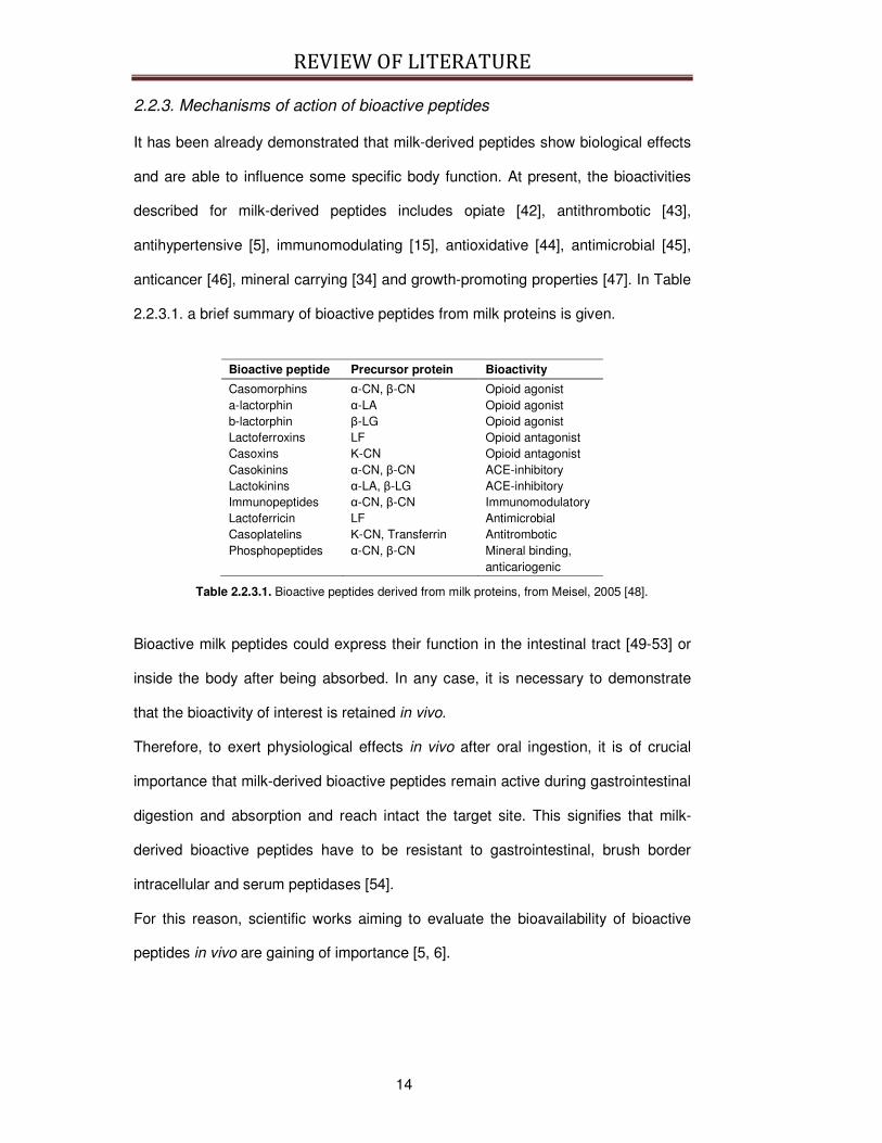
anticancer [46], mineral carrying [34] and growth-promoting properties [47]. In Table
2.2.3.1. a brief summary of bioactive peptides from milk proteins is given.
Bioactive peptide Precursor protein Bioactivity
Casomorphins α-CN, β-CN Opioid agonist a-lactorphin α-LA Opioid agonist b-lactorphin β-LG Opioid agonist Lactoferroxins LF Opioid antagonist Casoxins Κ-CN Opioid antagonist Casokinins α-CN, β-CN ACE-inhibitory Lactokinins α-LA, β-LG ACE-inhibitory Immunopeptides α-CN, β-CN Immunomodulatory Lactoferricin LF Antimicrobial Casoplatelins Κ-CN, Transferrin Antitrombotic Phosphopeptides α-CN, β-CN Mineral binding,
anticariogenic
Table 2.2.3.1. Bioactive peptides derived from milk proteins, from Meisel, 2005 [48].
Bioactive milk peptides could express their function in the intestinal tract [49-53] or
inside the body after being absorbed. In any case, it is necessary to demonstrate
that the bioactivity of interest is retained in vivo.
Therefore, to exert physiological effects in vivo after oral ingestion, it is of crucial
importance that milk-derived bioactive peptides remain active during gastrointestinal
digestion and absorption and reach intact the target site. This signifies that milk-
derived bioactive peptides have to be resistant to gastrointestinal, brush border
intracellular and serum peptidases [54].
For this reason, scientific works aiming to evaluate the bioavailability of bioactive
peptides in vivo are gaining of importance [5, 6].
REVIEW OF LITERATURE
15
2.2.4. Commercial dairy products and ingredients with health or function
claims based on bioactive peptides
It is now well documented that bioactive peptides can be generated during milk
fermentation with the starter cultures traditionally employed by the dairy industry. As
a result, peptides with various bioactivities can be found in the end-products, such
as various cheese varieties and fermented milks. These traditional dairy products
may, under certain conditions, carry specific health effects when ingested as part of
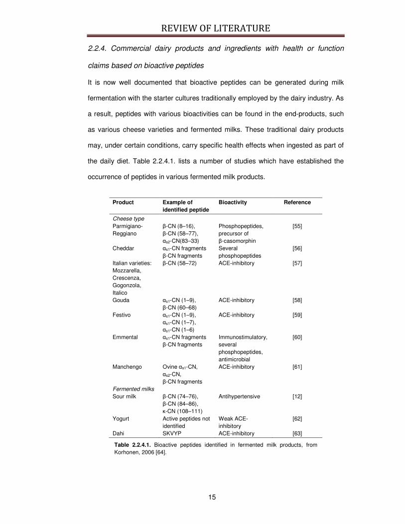
the daily diet. Table 2.2.4.1. lists a number of studies which have established the
occurrence of peptides in various fermented milk products.
Product Example of
identified peptide Bioactivity Reference
Cheese type Parmigiano-Reggiano
β-CN (8–16), β-CN (58–77), αs2-CN(83–33)
Phosphopeptides, precursor of β-casomorphin
[55]
Cheddar αs1-CN fragments β-CN fragments
Several phosphopeptides
[56]
Italian varieties: Mozzarella, Crescenza, Gogonzola, Italico
β-CN (58–72) ACE-inhibitory [57]
Gouda αs1-CN (1–9), β-CN (60–68)
ACE-inhibitory [58]
Festivo αs1-CN (1–9), αs1-CN (1–7), αs1-CN (1–6)
ACE-inhibitory [59]
Emmental αs1-CN fragments β-CN fragments
Immunostimulatory, several phosphopeptides, antimicrobial
[60]
Manchengo Ovine αs1-CN, αs2-CN, β-CN fragments
ACE-inhibitory [61]
Fermented milks Sour milk β-CN (74–76),
β-CN (84–86), κ-CN (108–111)
Antihypertensive [12]
Yogurt Active peptides not identified
Weak ACE-inhibitory
[62]
Dahi SKVYP ACE-inhibitory [63]
Table 2.2.4.1. Bioactive peptides identified in fermented milk products, from Korhonen, 2006 [64].
REVIEW OF LITERATURE
16
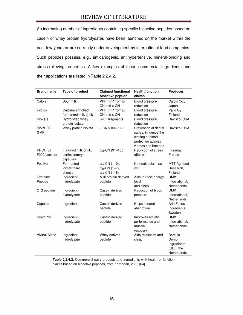
An increasing number of ingredients containing specific bioactive peptides based on
casein or whey protein hydrolysates have been launched on the market within the
past few years or are currently under development by international food companies.
Such peptides possess, e.g., anticariogenic, antihypertensive, mineral-binding and
stress-relieving properties. A few examples of these commercial ingredients and
their applications are listed in Table 2.2.4.2.
Brand name Type of product Claimed functional
bioactive peptide Health/function claims
Producer
Calpis Sour milk VPP, IPP from β-CN and κ-CN
Blood pressure reduction
Calpis Co., Japan
Evolus Calcium enriched fermented milk drink
VPP, IPP from β-CN and κ-CN
Blood pressure reduction
Valio Oy, Finland
BioZate Hydrolyzed whey protein isolate
β-LG fragments Blood pressure reduction
Davisco, USA
BioPURE-GMP
Whey protein isolate k-CN f(106–169)
Prevention of dental caries, influence the clotting of blood, protection against viruses and bacteria
Davisco, USA
PRODIET F200/Lactium
Flavored milk drink, confectionery, capsules
αs1-CN (91–100)
Reduction of stress effects
Ingredia, France
Festivo Fermented low-fat hard cheese
αs1-CN (1–9), αs1-CN (1–7), αs1-CN (1–6)
No health claim as yet
MTT Agrifood Research, Finland
Cysteine Peptide
Ingredient-hydrolysate
Milk protein derived peptide
Aids to raise energy level and sleep
DMV International, Netherlands
C12 peptide Ingredient-hydrolysate
Casein derived peptide
Reduction of blood pressure
DMV International, Netherlands
Capolac Ingredient Casein derived peptide
Helps mineral absorption
Arla Foods Ingredients, Sweden
PeptoPro Ingredient-hydrolysate
Casein derived peptide
Improves athletic performance and muscle recovery
DMV International, Netherlands
Vivinal Alpha Ingredient-hydrolysate
Whey derived peptide
Aids relaxation and sleep
Borculo Domo Ingredients (BDI), the Netherlands
Table 2.2.4.2. Commercial dairy products and ingredients with health or function claims based on bioactive peptides, from Korhonen, 2006 [64].
REVIEW OF LITERATURE
17
2.3. Bioactivities of interest
As already introduced in Paragraph 2.2.3., milk-derived bioactive peptides are
potential modulators of various regulatory processes in the body, and they can
express hormone-like activities.
Moreover, the primary sequence of some specific bovine proteins, as caseins,
contains overlapping regions, partially protected from proteolytic breakdown, that
manifest multifunctional properties and influence different biological functions [48]. In
particular, ACE-inhibitory and immunomodulatory properties seem to be associated,
possibly because both are correlated to the presence of short chain peptides such
as VPP and IPP formed during milk fermentation with selected bacterial stains [65].
Therefore, as the present thesis mainly focuses on milk-derived peptides displaying
immunomodulatory activity in the following paragraphs, immunomodulatory property
is described in more details together with the milk-derived peptides responsible for
these bioactivities. In addition, special attention is also given to ACE-inhibitory
activity and to the related bioactive peptides, because this activity can be associated
to immunomodulatory activity and because it has been the object of the Experiment
1 of this thesis.
2.3.1. ACE-inhibition
The inhibition of the Angiotensin-I-Converting Enzyme (ACE) is a key point in the
treatment of the hypertension. ACE is carboxypeptidase (E.C. 3.4.15.1) and
catalyzes the cleavage of dipeptides [66]. ACE is responsible for the conversion of
angiotensin I, a decapeptide generated by the action of rennin on the substrate
angiotensinogen, to the vasoconstrictor octapeptide angiotensin II. Angiotensin II
directly acts on blood vessels increasing blood pressure, but it also stimulates the
release of aldosterone from the adrenal cortex. Aldosterone increases the
reabsorption of sodium and water and the secretion of potassium by the kidney, so

REVIEW OF LITERATURE
18
the overall effect is an increased blood pressure (see Fig. 2.3.1.1.). In addition, ACE
hydrolyzes the vasodilatator bradykinin, inactivating its lowering pressure effects.
Fig. 2.3.1.1. Summarizing scheme of the effects of the rennin-angiotensin-aldosterone system, from http://en.wikipedia.org/wiki//File:Renin-angiotensin-aldosterone_system.png.
Human ACE is present into two isoforms, somatic ACE and germinal/testicular ACE.
Both isoforms are encoded by a single gene located on chromosome 17. The
somatic ACE is a membrane-bound protein expressed on the surface of the
vascular endothelial cells of the lungs and of the epithelial cells the kidney [67], but it
is widely distributes also in many other tissues as thymus and small intestine [68].
In some of these tissues the rennin–angiotensin-aldosterone system is not present:
this reinforces the idea that ACE has probably other roles in addition to the
production of angiotensin II and the inactivation of bradykinin.
2.3.1.1. Physiology of ACE-inhibition
Exogenous ACE-inhibitors having an antihypertensive effect in vivo were first
discovered in snake venom [69] and they are thought to be competitive substrates of
ACE. Indeed, the first ACE-inhibitor developed for the pharmacological treatment of

REVIEW OF LITERATURE
19
hypertension, Captopril, has been obtained modifying a peptide contained in the
venom of the a Brazilian snake [70] and designing it upon a hypothetical model of
the binding site on the enzyme [71]. Since then, synthetic ACE inhibitors such as
captopril, enalapril, alecepril and lisinopril are used extensively in the treatment of
essential hypertension despite their undesirable side effects, such as hypotension,
cough, increased potassium levels, reduced renal function, angioedema, etc. [33].
ACE-inhibitory peptides derived from milk proteins inhibit ACE as Captopril, thus
acting as competitive substrate of this enzyme, but they do not manifest the
correlated side effects [72].
Although the structure-activity relationship of ACE-inhibitory peptide has not been
fully elucidated, these peptides share common characteristics [73-75]:
• Short chain peptides (2-9 residues);
• Presence of hydrophobic residues in the sequence (aromatic or branched side
chains)
• Presence of proline, lysine or arginine residue at the C-terminal end of the
bioactive peptide
• Resistance to hydrolysis by digestive enzymes
Pripp and colleagues [76] established quantitative structure–activity relationships
(QSAR) for ACE-inhibitory peptides derived from milk proteins. For peptides up to
six amino acids, a relationship was found between the ACE-inhibitory activity and
some of the peptide characteristics (hydrophobicity and a positively charged amino
acid at the C-terminal position). No relationship was found between the N-terminal
structure and the ACE-inhibitory activity.
REVIEW OF LITERATURE
20
2.3.1.2. ACE-inhibitory peptides derived from milk
ACE-inhibitory peptides derived from milk proteins are released from caseins
(casokinins) or from whey proteins (lactokinins) [20, 48]. Casokinins and lactokinins
have been identified in fermented milks [77-79] or in milk proteins hydrolysates with
selected enzymes, such as pepsin, trypsin and chymotrypsin [40, 80-82].
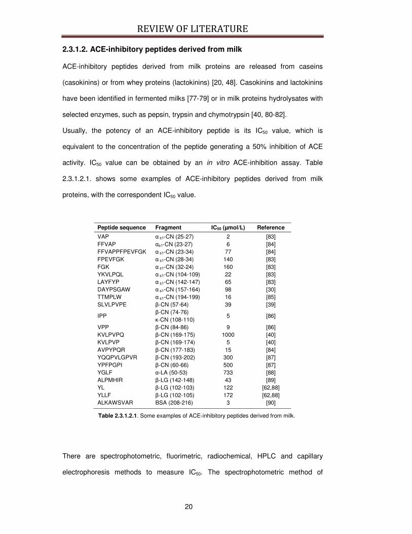
Usually, the potency of an ACE-inhibitory peptide is its IC50 value, which is
equivalent to the concentration of the peptide generating a 50% inhibition of ACE
activity. IC50 value can be obtained by an in vitro ACE-inhibition assay. Table
2.3.1.2.1. shows some examples of ACE-inhibitory peptides derived from milk
proteins, with the correspondent IC50 value.
Peptide sequence Fragment IC50 (µmol/L) Reference
VAP α s1-CN (25-27) 2 [83] FFVAP αs1-CN (23-27) 6 [84] FFVAPPFPEVFGK α s1-CN (23-34) 77 [84] FPEVFGK α s1-CN (28-34) 140 [83] FGK α s1-CN (32-24) 160 [83] YKVLPQL α s1-CN (104-109) 22 [83] LAYFYP α s1-CN (142-147) 65 [83] DAYPSGAW α s1-CN (157-164) 98 [30] TTMPLW α s1-CN (194-199) 16 [85] SLVLPVPE β-CN (57-64) 39 [39]
IPP β-CN (74-76) κ-CN (108-110)
5 [86]
VPP β-CN (84-86) 9 [86] KVLPVPQ β-CN (169-175) 1000 [40] KVLPVP β-CN (169-174) 5 [40] AVPYPQR β-CN (177-183) 15 [84] YQQPVLGPVR β-CN (193-202) 300 [87] YPFPGPI β-CN (60-66) 500 [87] YGLF α-LA (50-53) 733 [88] ALPMHIR β-LG (142-148) 43 [89] YL β-LG (102-103) 122 [62,88] YLLF β-LG (102-105) 172 [62,88] ALKAWSVAR BSA (208-216) 3 [90]
Table 2.3.1.2.1. Some examples of ACE-inhibitory peptides derived from milk.
There are spectrophotometric, fluorimetric, radiochemical, HPLC and capillary
electrophoresis methods to measure IC50. The spectrophotometric method of

REVIEW OF LITERATURE
21
Cushman and Cheung [91] is most commonly utilized. It is based on the hydrolysis
of hippuryl-His-Leu (HHL) by ACE to hippuric acid (HA) and HL. The extent of HA
release from HHL is measured after it is extracted with ethyl acetate. Direct,
extraction-free method has been published recently [92, 93]. Another broadly used
spectrophotometric method is based on the hydrolysis of a furanocryloyl tripeptide
(FA–Phe–Gly–Gly, FAPGG) to FAP and the dipeptide GG [94-96]. However, the
observation that the ACE-inhibitory activity differed with the method employed
creates a need to standardize the methodologies to evaluate in vitro ACE-inhibitory
activity [94, 95]. In practice, differences may arise among the results of different
assays due to the use of different substrates or, within the same assay, due to the
use of different test conditions or ACE from different origins. In particular, ACE
activity levels need to be carefully controlled to obtain comparable and reproducible
values [94, 96].
It has also to be considered that the IC50 value is not always directly related to the in
vivo hypotensive effects. Hypotensive effects can be measured in spontaneous
hypertensive rats (SHR), which are genetically predisposed to have a high blood
pressure and constitute an accepted model for human primary hypertension [72, 78,
79, 97-103] and in clinical trial with hypertensive patients [11, 82, 104, 105]. The
parameter monitored to assess the hypotensive effects of these products normally is
blood pressure in normal subjects or in subjects affected by hypertension [5], as
depicted in Figure 2.3.1.2.1..
Some peptides that manifest a reduced ACE-inhibitory activity in vitro express a
significant hypotensive effect when administered in vivo, confirming that the in vitro
ACE-inhibitory activity is not always directly related to the in vivo hypotensive
effects.

REVIEW OF LITERATURE
22
Fig. 2.3.1.2.1. Mean (±SEM) change in systolic blood pressure (SBP) and diastolic blood pressure (DBP) from baseline during the 21 weeks of treatment in the test product (; n = 19) and control (; n = 17) groups, from Seppo et al., 2003 [5].
For example, some milk-derived peptides have lower ACE-inhibitory activity in vitro
than the synthetic ACE inhibitor Captopril, but they usually display higher in vivo
activities than the efficacy levels extrapolated from the in vitro activities. This fact
has been attributed to a higher affinity to the tissues and a slower elimination [106],
but it may also be an indication of the existence of an additional mode of action than
the inhibition of ACE [54]. It could be possible that the peptides with a low in vitro
ACE-inhibitory activity could act as pro-drugs, releasing the active fragment by the
action of digestive or serum peptidases [97].
Conversely, some other ACE-inhibitory peptides manifest a high in vitro activity but
have no hypotensive effects in vivo. For example, the peptide FFVAP derived from
REVIEW OF LITERATURE
23
αs1-CN (23-27) is a potent ACE-inhibitory peptide in vitro (IC50 = 6 µmol/L) [84], but it
has not hypotensive effect in vivo.
The difficulty to establish a direct relationship between ACE-inhibitory activity in vitro
and antihypertensive activity in vivo may depend upon different reasons but peptide
bioavailability after oral administration plays a major role. As already introduced in
Paragraph 2.2.2., ACE-inhibitory peptides have to remain active during
gastrointestinal digestion and absorption and reach intact the target site.
As marked before, the evaluation of real hypotensive efficacy of peptides with high
in vitro ACE-inhibitory activity is further complicated by the different ACE-inhibition
assays that can be applied for the calculation of the IC50 [71, 94, 107]. In addition, in
in vivo experiment and clinical trials, different experimental designs (measurement
of arterial blood pressure at different points, different administration routes, or
doses) and the use of the animal model vs human experimentation make difficult to
examine the antihypertensive effects of ACE-inhibitory peptides.
However, testing the in vitro ACE-inhibitory activity could be still a necessary first
screening step, because it is based on a biological mechanism and the in vitro
assays are relatively easy and do not require expensive laboratory equipments.
Nevertheless, in vivo experiments and clinical trials are needed to demonstrate if the
hypotensive effect of these bioactive peptides is retained at physiological level.
Moreover, in vivo studies would permit to clarify the physiological mechanisms and
the targets of ACE-inhibitory peptides, once absorbed and circulating in the blood.
The main hypothesis on the mechanism of action of ACE-inhibitory milk peptides
assumes that absorbed peptides enter the blood circulation, concentrate in the aorta
where they exert their activity on the ACE expressed on the surface of endothelial
cells (Fig 2.3.1.2.2.) [5, 100].
REVIEW OF LITERATURE
24
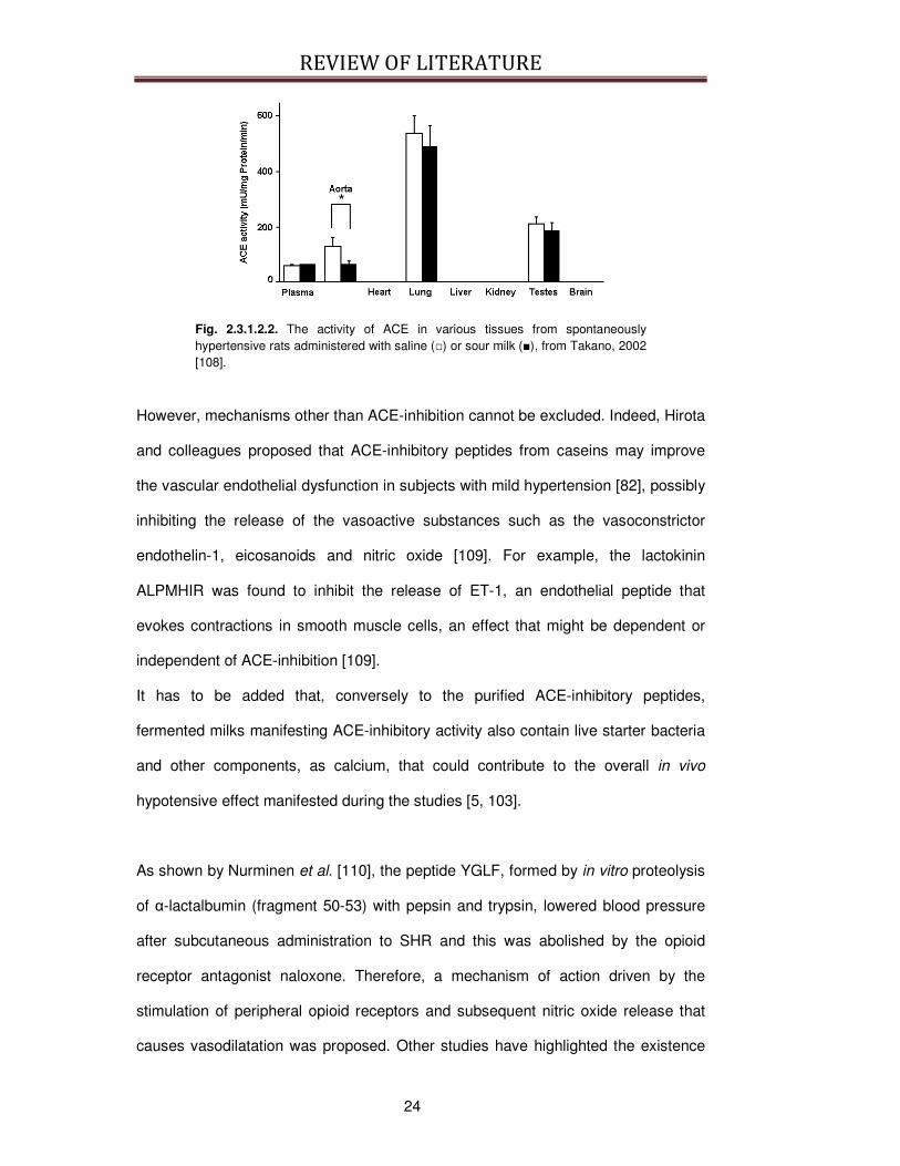
Fig. 2.3.1.2.2. The activity of ACE in various tissues from spontaneously hypertensive rats administered with saline () or sour milk (), from Takano, 2002 [108].
However, mechanisms other than ACE-inhibition cannot be excluded. Indeed, Hirota
and colleagues proposed that ACE-inhibitory peptides from caseins may improve
the vascular endothelial dysfunction in subjects with mild hypertension [82], possibly
inhibiting the release of the vasoactive substances such as the vasoconstrictor
endothelin-1, eicosanoids and nitric oxide [109]. For example, the lactokinin
ALPMHIR was found to inhibit the release of ET-1, an endothelial peptide that
evokes contractions in smooth muscle cells, an effect that might be dependent or
independent of ACE-inhibition [109].
It has to be added that, conversely to the purified ACE-inhibitory peptides,
fermented milks manifesting ACE-inhibitory activity also contain live starter bacteria
and other components, as calcium, that could contribute to the overall in vivo
hypotensive effect manifested during the studies [5, 103].
As shown by Nurminen et al. [110], the peptide YGLF, formed by in vitro proteolysis
of α-lactalbumin (fragment 50-53) with pepsin and trypsin, lowered blood pressure
after subcutaneous administration to SHR and this was abolished by the opioid
receptor antagonist naloxone. Therefore, a mechanism of action driven by the
stimulation of peripheral opioid receptors and subsequent nitric oxide release that
causes vasodilatation was proposed. Other studies have highlighted the existence
REVIEW OF LITERATURE
25
of vasorelaxant opioid peptides arising from β-Lactoglobulin such as β-LG (102-105)
[102] and from human casein, as YVPFPPF and YPFPPL [111].
2.3.1.3. Microorganisms and enzymes for the production of fermented
milk with ACE-inhibitory activity
At present, the in vivo hypotensive activity has been demonstrated for fermented
milks, milk proteins hydrolysates and purified ACE-inhibitory milk-derived peptides.
In vivo and in vitro studies have also confirmed that the microorganisms or the
peptidases used to obtain milk-derived products rich in ACE-inhibitory activity are of
extreme importance in influencing the quality and the quantity of ACE-inhibitory
peptides.
At the moment, the microorganisms used for the production of fermented milk with
ACE-inhibitory effects are selected for their high proteolytic activity and their food
safety, thus proteolytic LAB becoming the most used microorganisms. Nakamura
and colleagues [12] first selected a strain of L. helveticus together with
Saccharomyces cerevisiae to produce a fermented milk containing potent ACE-
inhibitory peptides, as IPP and VPP. Then L. helveticus has been preferred for the
purpose, although other LAB have shown good performance. Yogurt-type products
fermented with L. delb. bulgaricus and Lactobacillus lactis subsp. cremoris were
also shown to contain ACE-inhibitory peptides [29].
Recently, Muguerza et al. [112] assayed the ACE-inhibitory activity of fermented
milk samples produced with 231 microorganisms isolated from raw cow’s milk
samples. Among them, four E. faecalis strains resulted in the production of
fermented milk with potent ACE-inhibitory activity (IC50 = 34–59 µg/mL) and
antihypertensive activity in SHR.

REVIEW OF LITERATURE
26
Thus, strain selection is one of the main factors that influence the release of ACE-
inhibitory peptides. Tables 2.3.1.3.1a. and 2.3.1.3.1b. summarize the
microorganisms whose ability to produce fermented milks with high ACE-inhibitory
activity has been tested. However, further progress in this area may come from
elucidation of the specificity of microbial proteolytic systems in the integrated
environments prevailing in dairy products.
Microorganism Identified peptides Protein source Reference
L. helveticus CP790,
and S. cerevisiae
Nd β-CN, κ-CN [11, 39, 86]
L. helveticus LBK16H β-CN (74-76) κ-CN (108-110)
β-CN, κ-CN [5, 10, 103]
L. helveticus CPN4 β-CN (84-86) Whey proteins [113] L. helveticus NCC2765 β-CN (62-67)
β-CN (75-83) β-CN (149-153) β-CN (155-158) β-CN (183-190) β-CN (198-205) β-CN (208-213) β-CN (208-224) α s2-CN (205-212)
α s1-CN, β-CN [114]
L. helveticus CHCC637 Nd Milk [78] L. helveticus CHCC641 Nd Milk [78] Starter composed by a mix of S. thermophilus CR12,
L. casei LC01, L. helveticus PR4,
L. plantarum 1288
Nd Goat milk [115]
L. delb. bulgaricus SS1 β-CN (6-14) β-CN (7-14) β-CN (73-82) β-CN (74-82) β-CN (75-82)
β-CN, κ-CN [29]
L. lactis cremoris FT4 β-CN (6-14) β-CN (7-14) β-CN (47-52) β-CN (169-175) κ-CN (152-160) κ-CN (155-160)
β-CN, κ-CN [29]
K. marxianus marxianus β-LG [116] E. faecalis CECT5728 nd Bovine milk [79] E. faecalis CECT5727 nd Bovine milk [112] E. faecalis CECT5826 nd Bovine milk [112] E. faecalis CECT5827 nd Bovine milk [112]
Table 2.3.1.3.1a. Summary of microorganisms whose ability to produce fermented milk with high ACE-inhibitory activity has been tested. Nd: not determined.
REVIEW OF LITERATURE
27
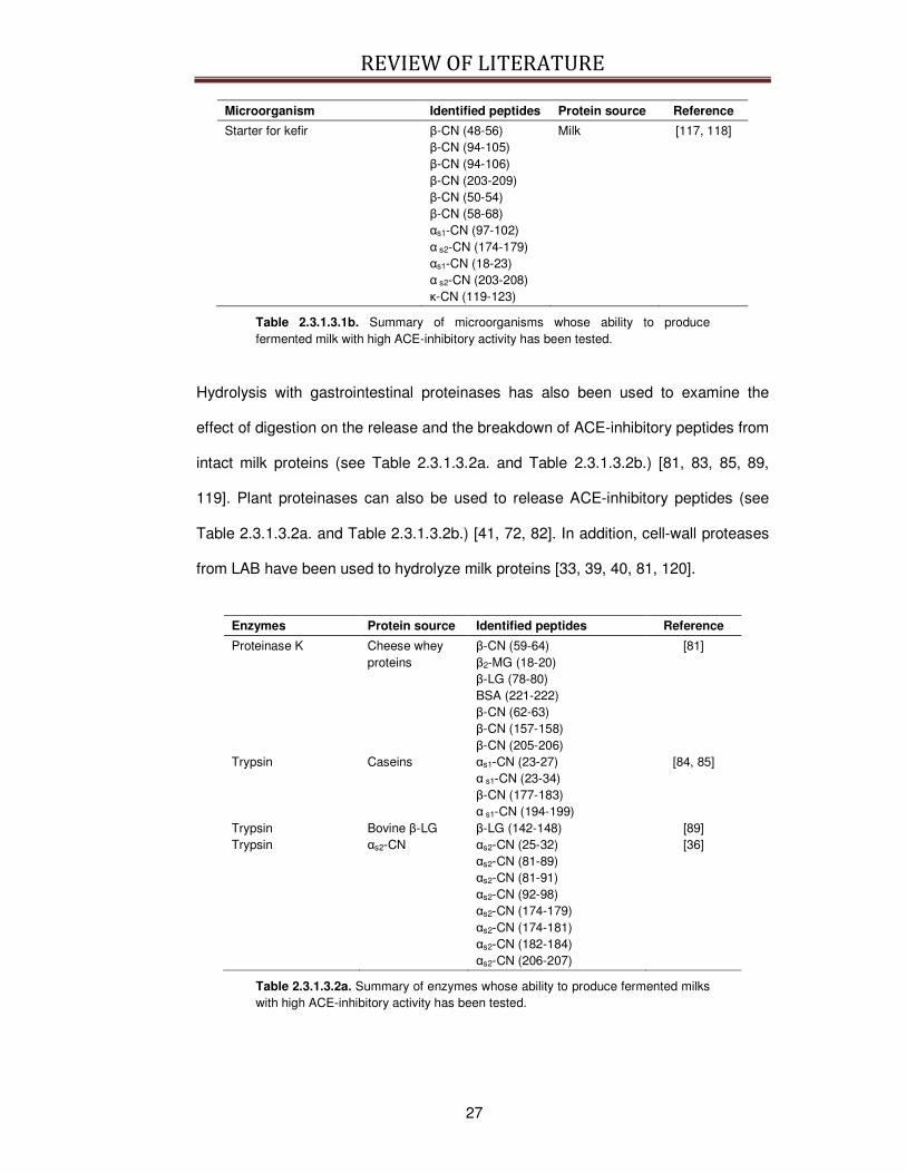
Microorganism Identified peptides Protein source Reference
Starter for kefir β-CN (48-56) β-CN (94-105) β-CN (94-106) β-CN (203-209) β-CN (50-54) β-CN (58-68) αs1-CN (97-102) α s2-CN (174-179) αs1-CN (18-23) α s2-CN (203-208) κ-CN (119-123)
Milk [117, 118]
Table 2.3.1.3.1b. Summary of microorganisms whose ability to produce fermented milk with high ACE-inhibitory activity has been tested.
Hydrolysis with gastrointestinal proteinases has also been used to examine the
effect of digestion on the release and the breakdown of ACE-inhibitory peptides from
intact milk proteins (see Table 2.3.1.3.2a. and Table 2.3.1.3.2b.) [81, 83, 85, 89,
119]. Plant proteinases can also be used to release ACE-inhibitory peptides (see
Table 2.3.1.3.2a. and Table 2.3.1.3.2b.) [41, 72, 82]. In addition, cell-wall proteases
from LAB have been used to hydrolyze milk proteins [33, 39, 40, 81, 120].
Enzymes Protein source Identified peptides Reference
Proteinase K Cheese whey proteins
β-CN (59-64) β2-MG (18-20) β-LG (78-80) BSA (221-222) β-CN (62-63) β-CN (157-158) β-CN (205-206)
[81]
Trypsin Caseins αs1-CN (23-27) α s1-CN (23-34) β-CN (177-183) α s1-CN (194-199)
[84, 85]
Trypsin Bovine β-LG β-LG (142-148) [89] Trypsin αs2-CN αs2-CN (25-32)
αs2-CN (81-89) αs2-CN (81-91) αs2-CN (92-98) αs2-CN (174-179) αs2-CN (174-181) αs2-CN (182-184) αs2-CN (206-207)
[36]
Table 2.3.1.3.2a. Summary of enzymes whose ability to produce fermented milks with high ACE-inhibitory activity has been tested.
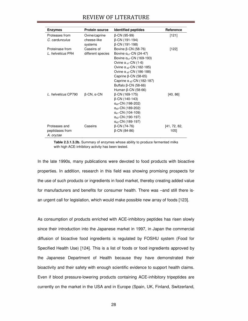
REVIEW OF LITERATURE
28
Enzymes Protein source Identified peptides Reference
Proteases from C. cardunculus
Ovine/caprine cheese-like systems
β-CN (95-99) β-CN (191-194) β-CN (191-198)
[121]
Proteinase from L. helveticus PR4
Caseins of different species
Bovine β-CN (58-76) Bovine αs1-CN (24-47) Bovine αs1-CN (169-193) Ovine α s1-CN (1-6) Ovine α s2-CN (182-185) Ovine α s2-CN (186-188) Caprine β-CN (58-65) Caprine α s2-CN (182-187) Buffalo β-CN (58-66) Human β-CN (58-66)
[122]
L. helveticus CP790 β-CN, α-CN β-CN (169-175) β-CN (140-143) αs2-CN (198-202) αs2-CN (189-202) αs1-CN (104-109) αs2-CN (190-197) αs2-CN (189-197)
[40, 86]
Proteases and peptidases from A. oryzae
Caseins β-CN (74-76) β-CN (84-86)
[41, 72, 82, 105]
Table 2.3.1.3.2b. Summary of enzymes whose ability to produce fermented milks with high ACE-inhibitory activity has been tested.
In the late 1990s, many publications were devoted to food products with bioactive
properties. In addition, research in this field was showing promising prospects for
the use of such products or ingredients in food market, thereby creating added value
for manufacturers and benefits for consumer health. There was –and still there is-
an urgent call for legislation, which would make possible new array of foods [123].
As consumption of products enriched with ACE-inhibitory peptides has risen slowly
since their introduction into the Japanese market in 1997, in Japan the commercial
diffusion of bioactive food ingredients is regulated by FOSHU system (Food for
Specified Health Use) [124]. This is a list of foods or food ingredients approved by
the Japanese Department of Health because they have demonstrated their
bioactivity and their safety with enough scientific evidence to support health claims.
Even if blood pressure-lowering products containing ACE-inhibitory tripeptides are
currently on the market in the USA and in Europe (Spain, UK, Finland, Switzerland,

REVIEW OF LITERATURE
29
Italy, Iceland and Portugal), there is not a regulatory framework for bioactive foods
or bioactive food ingredients. The rules to be applied are numerous and they
depend on the nature of the foodstuff. Actually, in Europe, the General Food Law
regulations definitively are applicable to regulate the use of bioactive peptides in the
food marked and the associate use of health claims [123].
The first beverage with ACE-inhibitory peptides was commercialized in Japan, with
the name Amiru S Calpis® (Calpis Co. Ltd., Japan). This fermented milk is produced
by fermenting milk with L. helveticus CP790 and S. cerevisiae. Nakamura et al. [12,
86], identified the peptides VPP and IPP and this beverage revealed a significant
decrease in systolic blood pressure when ingested by hypertensive men [10, 125].
A new milk drink launched by Unilever under the Flora/Becel pro-active® also
contains VPP and IPP. This product is the first European fermented milk drink
designed to help lowering blood pressure and it contains a casein hydrolysate
produced by A. oryzae protease and it has been marketed by Calpis as
AmealPeptide. Recently, a study [105] was conducted among patients with high-
normal blood pressure and mild hypertension, who took different doses of this
beverage and a significant difference in systolic blood pressure between the placebo
group and the group receiving the beverage was observed. In both cases, a higher
dose of VPP is necessary due to the lower potency of this peptide compared to IPP
[64, 125].
Another available commercial product is named Evolus® (Valio Ltd, Finland or
Kaiku Vitabrands, Spain), a fermented milk with L. helveticus LBK-16H, which also
exerts a significant antihypertensive effect in humans [5, 125, 126].

REVIEW OF LITERATURE
30
Other L. helveticus strains used in the production of antihypertensive fermented milk
foods are L. helveticus R211, R389 [127] and LMG 11474 [128], as well as
CHCC641 and CCCH637 from Chr. Hansen A/S [97].
Two other commercial products, a casein hydrolysate containing the peptide
FFVAPFEVFGK (as1-CN (23-34)) named Casein DP (Kanebo, Ltd, Japan), and C12
peptide (DMV, The Netherlands), and a whey protein hydrolysate (BioZate, Davisco,
US) were also claimed to lower blood pressure in humans [33, 129, 130].
2.3.2. Immunomodulation
The immune response can be influenced by various factors. Numerous reports
demonstrate that milk bioactive peptides can interact with the immune system at
different levels. The next paragraphs provide a brief overview of the immune system
and of the effects of the milk-derived peptides implicated in the modulation of
immune responses.
2.3.2.1. Overview of the physiology of the immune system
The immune system is a body wide network of cells, tissues, and organs that has
evolved to defend the body against pathogens and foreign material, generally called
as “non-self”. Pathogens include infectious organisms as bacteria, viruses and
parasites and foreign material include for example toxins. All the non-self
substances capable of triggering an immune response are known as antigens (from
the National Cancer Institute of USA,
www.cancer.gov/cancertopics/understandingcancer/immunesystem/).
The organs of the immune system are positioned throughout your body and include
the bone marrow that is involved in the production of the immune cells. The thymus,
where T lymphocytes mature; the spleen and the lymph nodes that contain
specialized compartments where immune cells gather and confront antigens.
REVIEW OF LITERATURE
31
In addition to these organs, clumps of lymphoid tissue are found in many parts of the
body, especially in the linings of the digestive tract (GALT), the airways (BALT) and
the various mucosal compartments of the body (MALT) (from the National Cancer
Institute of USA).
Cells of the immune system are of various nature and each population has a
particular role. Neutrophils are particularly active against bacteria. Monocytes
circulate in the bloodstream for about one to three days and then typically move into
tissues throughout the body, where they differentiate into tissue resident
macrophages or dendritic cells. Circulating monocytes are responsible for
phagocytosis of antigens. Basophils are granulocytic cells that release granules
containing histamine and they play a role in both parasitic infections and allergies.
Mast cells are very similar in morphology and function to basophils but they resident
cells of several types of tissues. Eosinophils are granulocites with the main role of
combating multicellular parasites and some infections. Finally, Natural killer cells (or
NK cells) are a type of cytotoxic lymphocyte. NK cells play a major role in the
rejection of tumors and cells infected by viruses. The cells kill by releasing the
proteins called perforin and granzyme that cause the target cell to die by apoptosis
(from http://en.wikipedia.org/wiki/Immune_system).
In particular, these cells populations constitute the first line defense against
antigens. In fact they are involved in the recruitment of the immune cells to sites of
infection, through the production of chemical factors. In addition they promote the
clearance of dead cell and they activate the process of inflammation that is one of
the first responses of the immune system to infection or irritation. The first response
to an antigen is rapid and important but it is not selective against the antigen and it
is called innate immune response. This means that the cells of the innate system
REVIEW OF LITERATURE
32
recognize and respond to pathogens in a generic way, not conferring long-lasting or
protective immunity to the host (from http://en.wikipedia.org/wiki/Immune_system).
Cooperating with the innate immune system to eliminate pathogens, the other part
of the immune system is the adaptative immune system that is composed of highly
specialized, systemic cells and processes that recognize and “remember” specific
pathogens. In this way the response to the pathogen is more selective and efficient
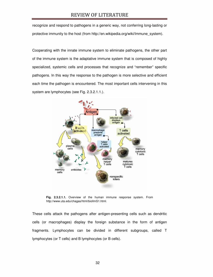
each time the pathogen is encountered. The most important cells intervening in this
system are lymphocytes (see Fig. 2.3.2.1.1.).
Fig. 2.3.2.1.1. Overview of the human immune response system. From http://www.uta.edu/chagas/html/biolImS1.html.
These cells attack the pathogens after antigen-presenting cells such as dendritic
cells (or macrophages) display the foreign substance in the form of antigen
fragments. Lymphocytes can be divided in different subgroups, called T
lymphocytes (or T cells) and B lymphocytes (or B cells).
REVIEW OF LITERATURE
33
The B cell turns into a plasma cell that produces and releases into the bloodstream
thousands of specific antibodies. Antibodies are large soluble proteins used to
recognize, identify and neutralize specific antigens. There are different types of
antibody, differing in biological properties, each has evolved to handle different kinds
of antigens (from http://en.wikipedia.org/wiki/Immune_system).
The T cells coordinate the entire immune response and eliminate the viruses hiding
in infected cells and contribute to the immune defenses in a cell-mediated way and
can be sub-grouped as follow. T helper cells (TH cells) assist other white blood cells
in immunologic processes, including maturation of B cells into plasma cells and
activation of cytotoxic T cells and macrophages, among other functions. Cytotoxic T
cells (TC cells, or CTLs) destroy virally infected cells and tumor cells, and are also
implicated in transplant rejection. After the infection has resolved, another subset of
antigen-specific T cells persist and they are called Memory T cells. They quickly
expand to large numbers of effector T cells upon re-exposure to their cognate
antigen, thus providing the immune system with "memory" against past infections.
Memory T cells comprise two subtypes: central memory T cells (TCM cells) and
effector memory T cells (TEM cells). Finally, Regulatory T cells (Treg cells), formerly
known as suppressor T cells, are crucial for the maintenance of immunological
tolerance. Their major role is to shut down T cell-mediated immunity toward the end
of an immune reaction and to suppress auto-reactive T cells that escaped the
process of negative selection in the thymus (from http://en.wikipedia.org/wiki/T_cell).
The efficient components of the immune system act cooperatively to eliminate the
infection. The “communication” between the different parts is mediated by
specialized chemical mediators, called cytokines. Cytokines are diverse and potent
chemical messengers secreted by the cells of the immune system. Cytokines
include interleukins, growth factors, and interferons.
REVIEW OF LITERATURE
34
Lymphocytes, including both T cells and B cells, secrete cytokines called
lymphokines, while the cytokines of monocytes and macrophages are called
monokines.
Many of these cytokines are also known as interleukins because they serve as a
messenger between leukocytes. Binding to specific receptors on target cells,
cytokines recruit many the different subsets of the immune system. In addition,
cytokines encourage cell growth, promote cell activation, direct cellular traffic, and
destroy target cells--including cancer cells. Moreover, it is common for different cell
types to secrete the same cytokine or for a single cytokine to act on several cell
types. Cytokines are redundant in their activity, meaning that the same function can
be stimulated by different cytokines.
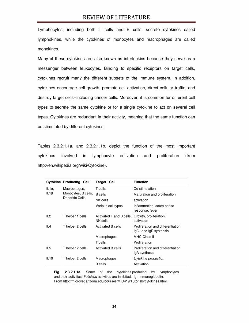
Tables 2.3.2.1.1a. and 2.3.2.1.1b. depict the function of the most important
cytokines involved in lymphocyte activation and proliferation (from
http://en.wikipedia.org/wiki/Cytokine).
Cytokine Producing Cell Target Cell Function
IL1α, IL1β
Macrophages, Monocytes, B cells, Dendritic Cells
T cells Co-stimulation
B cells Maturation and proliferation
NK cells activation
Various cell types Inflammation, acute phase response, fever
IL2 T helper 1 cells Activated T and B cells, NK cells
Growth, proliferation, activation
IL4 T helper 2 cells Activated B cells Proliferation and differentiation IgG1 and IgE synthesis
Macrophages MHC Class II
T cells Proliferation
IL5 T helper 2 cells Activated B cells Proliferation and differentiation IgA synthesis
IL10 T helper 2 cells Macrophages Cytokine production
B cells Activation
Fig. 2.3.2.1.1a. Some of the cytokines produced by lymphocytes and their activities. Italicized activities are inhibited. Ig: Immunoglobulin. From http://microvet.arizona.edu/courses/MIC419/Tutorials/cytokines.html.

REVIEW OF LITERATURE
35
Cytokine Producing Cell Target Cell Function
IL12 Macrophages B cells
Activated T cytotoxic cells
Differentiation into CTL (with IL2)
NK cells Activation
INFγ T helper 1 cells, T cytotoxic cells, NK cells
Various cell types Viral replication
Macrophages MHC expression
Activated B cells Ig class switch to IgG2a
T helper 2 cells Proliferation
Macrophages Pathogen elimination
TNFβ T helper 1 cells and T cytotoxic cells
Phagocytes Phagocytosis, NO production
Tumor cells Cell death
Fig. 2.3.2.1.1b. Some of the cytokines produced by lymphocytes and their activities. Italicized activities are inhibited. Ig: Immunoglobulin. From http://microvet.arizona.edu/courses/MIC419/Tutorials/cytokines.html.
In particular, two important cytokines involved in lymphocytes proliferation and
activation are IL2 and INFγ. IL2 is a T cell growth factor produced by T helper 1
(TH1) and NK cells. As an autocrine and paracrine growth factor, IL2 induces
proliferation and differentiation of T and B cells. IL2 is responsible for the progress of
T lymphocytes from the G1 to the S phase in the cell cycle and also for stimulation
of B cells for antibody synthesis. IL2 stimulates the growth of NK cells and enhances
the cytolytic function of these cells, producing lymphokine-activated killer cells. IL2
can also induce interferon INFγ secretion by NK cells.
INFγ is an important macrophage-activating lymphokine and it is involved in the
induction of other cytokines, particularly T Helper 2 cytokines, such as IL4, IL5, and
IL10. Because of its role in mediating macrophages and NK cell activation, INFγ is
important in host defense against intracellular pathogens and viruses and against
tumors responses thus influencing downstream immunological responses (From
http://microvet.arizona.edu/courses/MIC419/Tutorials/cytokines.html).
REVIEW OF LITERATURE
36
2.3.2.2. Immunomodulatory peptides derived from milk
Immunomodulatory milk peptides act on the immune system and cell proliferation
responses thus influencing downstream immunological responses and cellular
functions.
Indeed, in 1981 Jollés and colleagues [131] discovered that a tryptic hydrolysate of
human milk possessed in vitro immunostimulatory activity (more specifically,
stimulation of phagocytosis of sheep red blood cells and production of hemolytic
antibodies against the same cells).
In the following years, a number of potentially immunoregulatory peptides were
identified encrypted in bovine caseins [132-136] and whey proteins [137, 138], which
can manifest different effects (see Table 2.3.2.2.1a. and Table 2.3.2.2.1b.). Some
casein-derived peptides (residues 54-59 of human β-casein and residues 194-199
of αs1-casein) can stimulate phagocytosis of sheep red blood cells by murine
peritoneal macrophages [135, 139], exert a protective effect against Klebsiella
pneumoniae [140] or modulate proliferative responses and immunoglobulin
production in mouse spleen cell cultures (fragment 1-28 of bovine β-casein, [132,
141].
More recently, lactoferricin B, obtained by hydrolysis of lactoferricin by pepsin, was
found to promote phagocytic activity of human neutrophils [142]. Others fragments
(fragment 18-20 of κ-casein, fragment 90-96 of αs1-casein) can either stimulate or
inhibit lymphocyte proliferation depending upon the concentration used [134, 143],
while some whey-derived peptides can affect cytokine production from leucocytes
[137, 138].
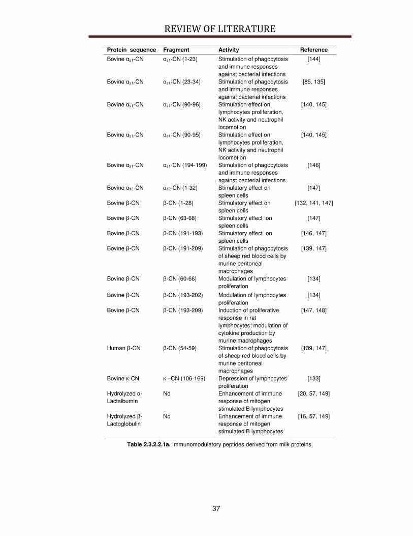
REVIEW OF LITERATURE
37
Protein sequence Fragment Activity Reference
Bovine αs1-CN αs1-CN (1-23) Stimulation of phagocytosis and immune responses against bacterial infections
[144]
Bovine αs1-CN αs1-CN (23-34) Stimulation of phagocytosis and immune responses against bacterial infections
[85, 135]
Bovine αs1-CN αs1-CN (90-96) Stimulation effect on lymphocytes proliferation, NK activity and neutrophil locomotion
[140, 145]
Bovine αs1-CN αs1-CN (90-95) Stimulation effect on lymphocytes proliferation, NK activity and neutrophil locomotion
[140, 145]
Bovine αs1-CN αs1-CN (194-199) Stimulation of phagocytosis and immune responses against bacterial infections
[146]
Bovine αs2-CN αs2-CN (1-32) Stimulatory effect on spleen cells
[147]
Bovine β-CN β-CN (1-28) Stimulatory effect on spleen cells
[132, 141, 147]
Bovine β-CN β-CN (63-68) Stimulatory effect on spleen cells
[147]
Bovine β-CN β-CN (191-193) Stimulatory effect on spleen cells
[146, 147]
Bovine β-CN β-CN (191-209) Stimulation of phagocytosis of sheep red blood cells by murine peritoneal macrophages
[139, 147]
Bovine β-CN β-CN (60-66) Modulation of lymphocytes proliferation
[134]
Bovine β-CN β-CN (193-202) Modulation of lymphocytes proliferation
[134]
Bovine β-CN β-CN (193-209) Induction of proliferative response in rat lymphocytes; modulation of cytokine production by murine macrophages
[147, 148]
Human β-CN β-CN (54-59) Stimulation of phagocytosis of sheep red blood cells by murine peritoneal macrophages
[139, 147]
Bovine κ-CN κ –CN (106-169) Depression of lymphocytes proliferation
[133]
Hydrolyzed α-Lactalbumin
Nd Enhancement of immune response of mitogen stimulated B lymphocytes
[20, 57, 149]
Hydrolyzed β-Lactoglobulin
Nd Enhancement of immune response of mitogen stimulated B lymphocytes
[16, 57, 149]
Table 2.3.2.2.1a. Immunomodulatory peptides derived from milk proteins.
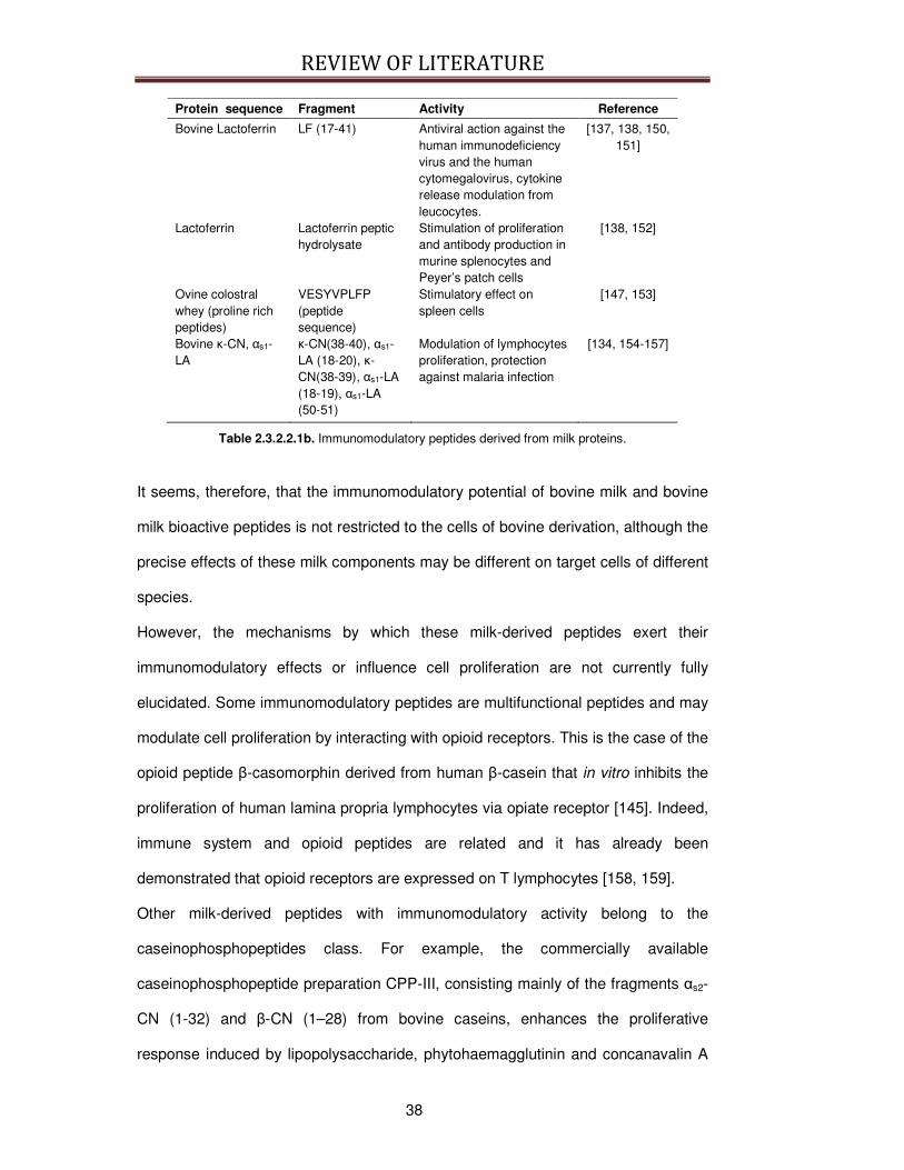
REVIEW OF LITERATURE
38
Protein sequence Fragment Activity Reference
Bovine Lactoferrin LF (17-41) Antiviral action against the human immunodeficiency virus and the human cytomegalovirus, cytokine release modulation from leucocytes.
[137, 138, 150, 151]
Lactoferrin Lactoferrin peptic hydrolysate
Stimulation of proliferation and antibody production in murine splenocytes and Peyer’s patch cells
[138, 152]
Ovine colostral whey (proline rich peptides)
VESYVPLFP (peptide sequence)
Stimulatory effect on spleen cells
[147, 153]
Bovine κ-CN, αs1-LA
κ-CN(38-40), αs1-LA (18-20), κ-CN(38-39), αs1-LA (18-19), αs1-LA (50-51)
Modulation of lymphocytes proliferation, protection against malaria infection
[134, 154-157]
Table 2.3.2.2.1b. Immunomodulatory peptides derived from milk proteins.
It seems, therefore, that the immunomodulatory potential of bovine milk and bovine
milk bioactive peptides is not restricted to the cells of bovine derivation, although the
precise effects of these milk components may be different on target cells of different
species.
However, the mechanisms by which these milk-derived peptides exert their
immunomodulatory effects or influence cell proliferation are not currently fully
elucidated. Some immunomodulatory peptides are multifunctional peptides and may
modulate cell proliferation by interacting with opioid receptors. This is the case of the
opioid peptide β-casomorphin derived from human β-casein that in vitro inhibits the
proliferation of human lamina propria lymphocytes via opiate receptor [145]. Indeed,
immune system and opioid peptides are related and it has already been
demonstrated that opioid receptors are expressed on T lymphocytes [158, 159].
Other milk-derived peptides with immunomodulatory activity belong to the
caseinophosphopeptides class. For example, the commercially available
caseinophosphopeptide preparation CPP-III, consisting mainly of the fragments αs2-
CN (1-32) and β-CN (1–28) from bovine caseins, enhances the proliferative
response induced by lipopolysaccharide, phytohaemagglutinin and concanavalin A
REVIEW OF LITERATURE
39
(conA) stimulation, and immunoglobulin production in mouse spleen cell cultures
[14, 160]; this immunostimulating activity was attributed to the o-phospho-L-serine
residue, hence suggesting that such a bioactivity is relatively stable to proteinase
action in the intestinal tract [161]. The study of Otani et al. [162] focusing on the
effects of CPP-III on serum and intestinal immunoglobulin G and immunoglobulin A
secretion in mice proved that oral use of caseinophosphopeptide is beneficial toward
enhancement of the mucosal immunity.
In addition, an alternative hypothesis involves a possible immunomodulatory action
via ACE-inhibitory mechanism. ACE-inhibitory peptides are well known for their
antihypertensive properties because they inhibit the conversion of angiotensin I to
angiotensin II, but have also been found to prevent cleavage of bradykinin, that is
mediated by ACE [135]. Bradykinin acts as a mediator of the acute inflammatory
process and is thus able to stimulate macrophages, enhance lymphocyte migration
and induce the secretion of cytokines from lymphocyte in culture. It should be noted
that a common structural feature of several ACE-inhibitory peptides and some
immunomodulatory peptides is the presence of arginine as the C-terminal residue
[163].
Immunomodulatory milk-derived peptides may contribute to the overall immune
response and may ameliorate immune system function. Migliore-Samour [140]
suggested that casein derived peptides are involved in the stimulation of the
newborn’s immune system. It cannot be excluded that the immunostimulating
activities may also have a direct effect on the resistance to bacterial and viral
infection of adult humans.
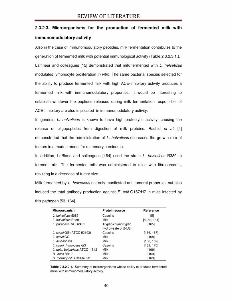
REVIEW OF LITERATURE
40
2.3.2.3. Microorganisms for the production of fermented milk with
immunomodulatory activity
Also in the case of immunomodulatory peptides, milk fermentation contributes to the
generation of fermented milk with potential immunological activity (Table 2.3.2.3.1.).
Laffineur and colleagues [15] demonstrated that milk fermented with L. helveticus
modulates lymphocyte proliferation in vitro. The same bacterial species selected for
the ability to produce fermented milk with high ACE-inhibitory activity produces a
fermented milk with immunomodulatory properties. It would be interesting to
establish whatever the peptides released during milk fermentation responsible of
ACE-inhibitory are also implicated in immunomodulatory activity.
In general, L. helveticus is known to have high proteolytic activity, causing the
release of oligopeptides from digestion of milk proteins. Rachid et al. [4]
demonstrated that the administration of L. helveticus decreases the growth rate of
tumors in a murine model for mammary carcinoma.
In addition, LeBlanc and colleagues [164] used the strain L. helveticus R389 to
ferment milk. The fermented milk was administered to mice with fibrosarcoma,
resulting in a decrease of tumor size.
Milk fermented by L. helveticus not only manifested anti-tumoral properties but also
induced the total antibody production against E. coli O157:H7 in mice infected by
this pathogen [53, 164].
Microorganism Protein source Reference
L. helveticus 5089 Caseins [15] L. helveticus R389 Milk [4, 52, 164] L. paracasei NCC2461 Tryptic-chymotryptic
hydrolysate of β-LG [165]
L. casei GG (ATCC 53103) Caseins [166, 167] L. casei GG Milk [168] L. acidophilus Milk [168, 169] L. casei rhamnosus GG Caseins [169, 170] L. delb. bulgaricus ATCC11842 Milk [169] B. lactis BB12 Milk [169] S. thermophilus DSM4022 Milk [169]
Table 2.3.2.2.1. Summary of microorganisms whose ability to produce fermented milks with immunomodulatory activity.
REVIEW OF LITERATURE
41
Fermented milks with immunomodulatory properties are not produced exclusively by
L. helveticus (Table 2.3.2.2.1.). Milk fermented by L. paracasei [165] was shown to
produce peptides from β-lactoglobulin that stimulate IL10 production and depress
lymphocyte proliferation. Additionally, L. casei GG was used to produce a casein
hydrolysate that suppresses human T cell activation, modulating IL2 expression
[166, 167, 170].
The immunomodulatory activity is independent from the presence of living
microorganisms, as evidenced by Perdigon [168] and by Vinderola [52] who
reported that the supernatant of fermented milk cultured with L. casei, L. acidophilus
and L. helveticus strains increased the immune response independently from the
presence of lactobacilli. This result was obtained also by De Simone [13] that tested
the INFγ production of human peripheral blood lymphocytes in response to filtered
yoghurt devoid of microorganisms. More recently LeBlanc examined the antibody
production following E. coli O157:H7 infection following the administration of a cell-
free supernatant from L. helveticus fermented milk and found that the increased
antibody production is not related to viable microorganism [53].
Microorganisms other that bacteria, as a cell-free extract obtained from the yeast S.
cerevisiae can be used for milk fermentation, producing a milk hydrolysate with
potential apoptosis-inducing effect in human leukemia HL-60 cells, as observed by
Roy et al. [171].
In addition, as already demonstrated for milk proteins [172, 173], bioactive peptides
present in yoghurt actually decreased cell proliferation with IEC-6 or Caco-2 cells,
which may explain, at least partially, why consumption of yoghurt has been
associated with a reduced incidence of colon cancer [174, 175].
The molecular mechanism by which the previous mentioned microorganisms
enhance the immune system is not yet clear but the previously discussed reports

REVIEW OF LITERATURE
42
strongly support the fact that immunomodulatory peptides released in fermented
milk contribute to the immunoenhancing and antitumor properties of dairy products.
It should be stressed that the extreme difficulty to establish how immunomodulatory
peptides and fermented milks influence the immune function is strictly linked to the
immune system complexity. Paragraph 2.3.2.1. already demonstrated that this
system comprises a complex interplay between different cell populations and
molecules. Thus, when the immunomodulatory activity of a bioactive peptide is
assessed in vitro, the single experimental result could demonstrate the specific
involvement of a particular milk-derived peptide in an immune mechanism but this
result is not conclusive in determining if this peptide its effects would be significant
for the whole immune system. For this reason the preferred term to describe the
influence of milk on the immune system is modulation, because the potential for
enhancement and suppression depends also on the target cells chosen to test the
immunomodulatory activity of the bioactive peptide [1].
2.3.2.4. Two examples of immunomodulatory peptides derived from
milk proteins
At present, most attention on immunomodulatory peptides has been focused on
lactoferricin, a pepsin-derived peptide from lactoferrin [132, 176-178], and on
glycomacropeptide, a k-casein-derived peptide (κ-CN (106–169)) [179, 180] present
in appreciable amounts in some whey protein concentrates and whey protein
isolates. But, as already revised before, during milk fermentation a number of
bioactive peptides are formed by enzymatic hydrolysis of milk proteins with
immunomodulatory potential. Particular attention will be given in this PhD thesis to
the fragment α-LA (18-20) (a tri-peptide named YGG) and to the long fragment β-CN
(193-209) because they have been chosen as model peptides to study the
immunomodulatory activity and the absorption mechanism of bioactive peptides
derived from milk proteins.
REVIEW OF LITERATURE
43
2.3.2.4.1. YGG peptide
The peptide YGG (Tyr-Gly-Gly) represents an interesting example of cryptic peptide
with putative immunomodulating effects, as it can originate from at least two
different sources. First, it is the product of the hydrolysis of Leu-enkephalin and Met-
enkephalin [181, 182], and thus it is an endogenous peptide. In addition, it can be
considered as a potential nutraceutical, because it is also encrypted in milk proteins
and can be released during the digestion of bovine milk, in particular from α-
lactalbumin (fragment 18-20) [31, 134].
It is known that Met-enkephalins, the YGG endogenous progenitor, can enhance
human T cell proliferation and IL2 production in vitro in the absence of mitogens,
possibly through the activation of opioid receptors present on the cell surface [183].
The enhancement of human peripheral blood lymphocytes proliferation and protein
synthesis in vitro was obtained also with YGG administration in presence of conA
[134, 184]. In addition, it was observed that YGG can affect INFγ and IL2 secretion
in murine splenocytes stimulated with suboptimal concentration of conA in serum-
free medium [157].
Stimulatory effects on cell proliferation were observed also in leukocytes obtained
from mice administrated in vivo with either Met-enkephalin or YGG, suggesting that
Met-enkephalin effects on the immune cells are mediated by YGG [185]. More
recently, the immunomodulatory effect of YGG was confirmed in vivo by the
observation that the peptide administration modulated the delayed-type
hypersensitivity responses to tuberculin derivatives in hairless guinea pigs [154]. It is
noteworthy to observe that YGG seems to have a biphasic effect on the parameters
studied so far, as it showed an enhancing effect at low doses and an inhibitory effect
at higher doses [154, 157].
It should be noted that YGG is contained several times in the primary structure of
bovine κ-casein and α-lactalbumin and it could be released during milk fermentation
or gastrointestinal digestion from the precursor proteins. In addition, it is a tripeptide
REVIEW OF LITERATURE
44
and, as already demonstrated for other milk-derived bioactive peptides [186], it can
be assumed that it can pass across the intestine by a carrier-mediated peptide
transport system in quantitatively significant amounts and, hence, may reach
peripheral target sites.
2.3.2.4.2. β-CN (193-209) peptide
The β-CN (193–209) peptide is released from the C-terminal end of β-casein by
hydrolysis with pepsin-chymosin. It is a 17 residues long peptide with the amino acid
sequence Tyr-Gln-Glu-Pro-Val-Leu-Gly-Pro-Val-Arg-Gly-Pro-Phe-Pro-Ile-Ile-Val.
This peptide was isolated and identified from yoghurt and fermented milks as well as
several types of cheese including Feta and Camembert studies [187, 188].
This peptide displays immunomodulatory properties and shows mitogenic activity on
primed lymph node cells and unprimed rat spleen cells [147], it manifests
chemotactive activity on L14 lymphoblastoid cell line [189], and enhances
phagocytosis in rat macrophages [148, 190].
In addition, a smaller fragment of β-CN (193–209), corresponding to the amino acid
sequence Gly-Pro-Val-Arg-Gly-Pro-Phe-Pro-Ile-Ile, displayed ACE-inhibitory activity,
further supporting the concept that ACE-inhibitors may also act as
immunomodulatory peptides by acting as bradykinin-potentiating peptides [37].
Interestingly, the presence of 4 proline residues within the sequence can protect the
long peptide β-CN (193–209) from the action of peptidases. So it could be possible
that this peptide can cross the intestinal barrier in an intact bioactive form.
2.4. Bioactive peptide digestion
Some bioactive peptides can express their activity directly on the gastrointestinal
tract but the majority of them has to reach their target site inside the body. They
have to remain stable during the digestion process and cross the gastrointestinal

REVIEW OF LITERATURE
45
barrier maintaining their biological activities. It is thus important to know the
physiology of digestion of proteins and peptides in the gastrointestinal tract, more
specifically the human GI system, to understand the mechanisms determining the
bioavailability of bioactive peptides in vivo.
2.4.1. Physiology of the digestion of proteins and peptides
In humans, the most important sites for the digestion of proteins and peptides are
the stomach and the small intestine. The stomach is the portion of the GI tract that is
located between the cardia and pylorus valves (see Fig. 2.4.1.1.). It can be divided
in different regions which differ for the structure and functionality of the glands
distributed in the gastric mucosa.
Fig. 2.4.1.1. The anatomic structure of the human stomach, from www.acm.uiuc.edu/sigbio/project/digestive/middle/stomach2.jpg
The gastric glands are composed by different types of cells, as HCl-secreting
parietal cells, pepsinogen-secreting cells, mucous-secreting cells, and endocrine
cells (see Fig. 2.4.1.2.).

REVIEW OF LITERATURE
46
Fig. 2.4.1.2. Structure and function of a gastric gland, from www.colorado.edo/intphys/class/IPHY3430-200/image/21-25.jpg
The human small intestine is 2–6 m in length and is loosely divided into three
sections – duodenum, jejunum and ileum – which comprise 5%, 50% and 45% of
the length, respectively (see Fig. 2.4.1.3.).
Fig. 2.4.1.3. The three sections of the small intestine, that is duodenum, jejunum (rose) and ileum (yellow), from www.yoursurgery.com/procedures/intussussception/images/SmBowelAnat.jpg.

REVIEW OF LITERATURE
47
The surface of the small intestinal region has various unique projections that
significantly increase the potential surface area available for digestion and
absorption. Macroscopic valve-like folds encircle the inside of the intestinal lumen
and increase the surface area of the small intestine threefold. In addition, the
presence of villi and microvilli increases the surface area by 30-fold and 600-fold,
respectively. In particular, brush border membrane is the highly folded membrane
that covers the entire surface of the small intestine and constitutes the massive
surface area cited earlier. It is highly developed as a metabolically functional
membrane, incorporating a selection of enzymes, transporters and receptors [191].
The key function of the small intestine is the selective absorption of major nutrients.
In addition, it serves as a barrier to digestive enzymes and ingested foreign
substances.
Fig. 2.4.1.4. The structure of lumen and of the epithelium of the small intestine, from http://kvhs.nbed.nb.ca/gallant/biology/small_intestine.jpg
The epithelial cells in the intestinal region are of heterogeneous nature, and they
include enterocytes or absorptive cells, goblet cells (that secrete mucin), endocrine
cells, Paneth cells, M cells and tuft and cup cells (see Fig. 2.4.1.4.). Enterocytes are
the most common epithelial cells and they are thus responsible for the majority of
the absorption of nutrients and drugs in the small intestine. The enterocytes are
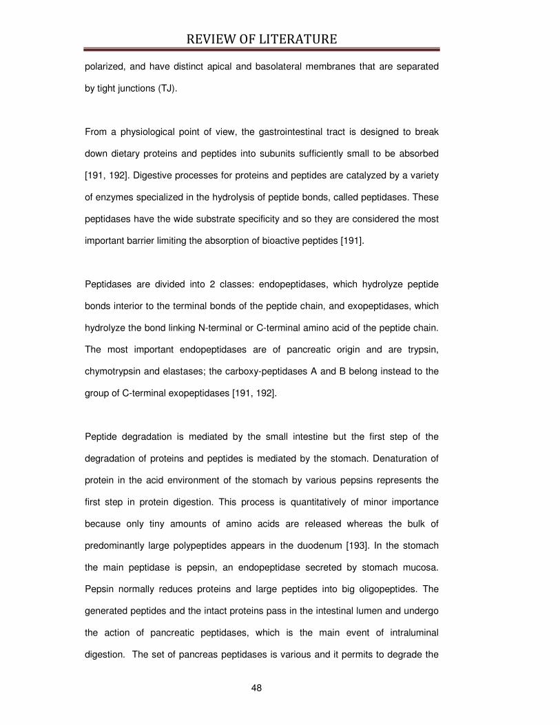
REVIEW OF LITERATURE
48
polarized, and have distinct apical and basolateral membranes that are separated
by tight junctions (TJ).
From a physiological point of view, the gastrointestinal tract is designed to break
down dietary proteins and peptides into subunits sufficiently small to be absorbed
[191, 192]. Digestive processes for proteins and peptides are catalyzed by a variety
of enzymes specialized in the hydrolysis of peptide bonds, called peptidases. These
peptidases have the wide substrate specificity and so they are considered the most
important barrier limiting the absorption of bioactive peptides [191].
Peptidases are divided into 2 classes: endopeptidases, which hydrolyze peptide
bonds interior to the terminal bonds of the peptide chain, and exopeptidases, which
hydrolyze the bond linking N-terminal or C-terminal amino acid of the peptide chain.
The most important endopeptidases are of pancreatic origin and are trypsin,
chymotrypsin and elastases; the carboxy-peptidases A and B belong instead to the
group of C-terminal exopeptidases [191, 192].
Peptide degradation is mediated by the small intestine but the first step of the
degradation of proteins and peptides is mediated by the stomach. Denaturation of
protein in the acid environment of the stomach by various pepsins represents the
first step in protein digestion. This process is quantitatively of minor importance
because only tiny amounts of amino acids are released whereas the bulk of
predominantly large polypeptides appears in the duodenum [193]. In the stomach
the main peptidase is pepsin, an endopeptidase secreted by stomach mucosa.
Pepsin normally reduces proteins and large peptides into big oligopeptides. The
generated peptides and the intact proteins pass in the intestinal lumen and undergo
the action of pancreatic peptidases, which is the main event of intraluminal
digestion. The set of pancreas peptidases is various and it permits to degrade the

REVIEW OF LITERATURE
49
majority of large peptides. Most of these enzymes are secreted by the pancreas in
an inactive form (trypsinogen, chymotrysinogen, proelastase, procarboxypeptidase).
Activation of trypsinogen requires enterokinase, a small intestinal mucosal enzyme.
Activation of the other precursors requires trypsin. Intraluminal hydrolysis of large
polypeptides results in oligopeptides composed of 2-8 amino acids. The luminal
phase of protein digestion leads therefore mainly to appearance of oligopeptides,
but only to small amounts of free amino acids [193].
The relative importance of this luminal hydrolysis in the overall degradation is
dependent on the size and the respective amino acid composition of the peptide
[194]. However, even when luminal peptide degradation occurs, it constitutes at best
the 20% of the total degradation in a given intestinal segment. This implies that
significant degradation of the peptide requires at least the contact with brush border
membrane or uptake into the enterocytes [192]. Indeed, peptidases in the brush
border membrane are probably the biggest deterrent to the absorption of small
peptides across the intestinal mucosa [194] and the mucosa of small intestine
expresses at least 15 peptidases (see Table 2.4.1.1a. and Table 2.4.1.1b.).
Enzyme Substrates and Properties Products
Endopeptidases: Hydrolysis of internal peptide bonds: Enterokinase - Of trypsinogen (initiation of luminal
digestion) Trypsin
Neutral endopeptidase (EC 3.4.24.11)
- At hydrophobic amino acids of α-casein, insulin etc.
Peptides
PABA peptide hydrolase
Dipeptidases: Hydrolysis of dipeptides into AA Zn-stable Asp-Leu peptidase Dipeptides, esp. Asp-Leu Amino acids Gly-Leu peptidase Dipeptides, esp. Gly-Leu metalloenzyme
(Zn) Amino acids
Membrane dipeptidases Dipeptides (glutathione conjugate) Amino acids
Table 2.4.1.1a. Intestinal brush border associated proteases and peptidases, from Hartmann et al., 2007 [195].

REVIEW OF LITERATURE
50
Enzyme Substrates and Properties Products
Oligopeptidases: Hydrolysis of N-terminal peptide bonds:
Aminopeptidases (AP) AP N (EC 3.4.11.2) AP A (EC 3.4.11.7) AP P (EC 3.4.11.9) AP W (EC 3.4.11.16)
AP are metalloenzymes (Zn, Ca, Co). AA-oligopeptide of neutral amino acids AA-oligopeptide of acidic amino acids Pro-oligopeptide X-Trp, X-Trp-oligopeptide
Amino acids di-, tripeptides
Dipeptidyl-AP IV (DPP IV; EC 3.4.14.5)
X-Pro-oligopeptide X-Ala-oligopeptide X-Lys-oligopeptide DPP IV is a serine protease.
X-Pro, X-Ala, X-Lys, oligo-, dipeptides
Carboxypeptidases (CP): Hydrolysis of C-terminal peptide bonds: dipeptidyl-CP I; EC 3.4.15.1 Angiotensin,
Oligopeptide-Pro Synonym: angiotensin-converting enzyme, peptidase P
Peptides
γ-glutamyl-transpeptidase EC 2.3.2.2
Peptides with bound γ-glutamyl, e.g. gluthation
Peptides, γ -glutamyl-AA
Folate conjugase; EC 3.4.19.9 Polyglutamyl folate Folic acid CP M; EC 3.4.17.12 Peptide-Lys, peptide-Arg Basic amino
acids
Table 2.4.1.1b. Intestinal brush border associated proteases and peptidases, from Hartmann et al., 2007 [195].
In general it appears that brush-border peptidases are active mainly against tri-,
tetra, and higher peptides reducing them to the amino acid residues [192, 196] (see
Fig. 2.4.1.5.). The cytosol indeed contains a set of peptidases particularly active
against di and tri peptides. So, after the action of brush border peptidases, there is
only little possibility that large peptides could be absorbed.
Regardless of the mechanisms of absorption, the bioactive peptides that enter the
enterocyte undergo the action of the peptidases of the cytosol or the cellular
organelles. Indeed, the lysosome contains a massive array of enzymes, estimated
over 60 in number, which are capable of degrading any biological macromolecule,
including peptides and proteins [194, 197].
REVIEW OF LITERATURE
51
Fig. 2.4.1.5. Schematic representation of the brush border membrane and cytoplasmic phase of intestinal protein assimilation. The dual mechanism of peptide absorption is indicated in the dashed line. AOP = amino-oligopeptidase, N-AA-carrier = carrier for neutral amino acids, B-AA-carrier = carrier for basic amino acids. From Caspary, 1992 [193].
2.4.1.1. The digestion of bioactive peptides derived from milk proteins
The release of ACE-inhibitory peptides upon gastrointestinal digestion of milk
proteins or protein fragments, as well as the resistance to digestion of known ACE-
inhibitory sequences has been tested in several in vitro studies where the
gastrointestinal process was mimicked by the sequential hydrolysis with pepsin and
pancreatic enzymes (trypsin, chymotrypsin, carboxy and aminopeptidases). These
studies showed that gastrointestinal digestion is an essential factor in determining
ACE-inhibitory activity [188, 198]. The conditions of the simulated gastrointestinal
digestion (enzyme preparation, temperature, pH and incubation time) greatly
influence the degree of proteolysis and the resultant ACE-inhibitory activity [198].
The action of brush-border peptidases, the recognition by intestinal peptide
transporters and the subsequent susceptibility to plasma peptidases also determine
the physiological effect [54].
REVIEW OF LITERATURE
52
2.4.2. Digestion Models
Resistance to hydrolysis is one of the main factors influencing the bioavailability of a
bioactive peptide. However, few in vitro and ex vivo models are available about the
simulation of the digestion of bioactive peptides. In addition, the majority of the
reports are not specifically realized to study the digestion of bioactive peptides but
their main purpose is to develop digestion models for the evaluation of potential
allergenicity of food proteins [199], in particular milk proteins [200]. On the contrary,
a consistent body of literature exists about in vitro and ex vivo models for the
absorption of bioactive peptides, as Caco-2 cell lines [186, 201, 202].
The effects of digestive enzymes on bioactive peptides, in particular ACE-inhibitory
peptides derived from different food matrices, have been evaluated in in vitro
gastrointestinal simulated systems [203]. The first methods were developed by
Garrett and colleagues on soy proteins [204] and then slightly modified by
Hernandez-Ledesma and colleagues [188], Picariello and colleagues [205] and Lo
and colleagues [206] that applied the protocol on milk proteins. The common
purpose of these experiments was to assess the effects of the peptidases of the
stomach and the pancreas on the preservation of the ACE-inhibitory activity of
different hydrolysates. Other authors [207, 208] preferred to use the Corolase PP®
instead of the pancreatin. The Corolase PP® is a proteolytic enzyme preparation
from pig pancreas glands that contains, in addition to trypsin and chymotrypsin,
numerous amino-acid- and carboxy-peptidases.
However, these models are not completely predictive of the resistance of the
bioactive peptides because they do not mimic all the physiological factors affecting
food digestion, as pH variations, the relative amounts of the enzymes, the
interactions with other molecules, and the ratio peptidase/tested compound. These
variations may affect the rate of enzymatic degradation of the bioactive peptides
REVIEW OF LITERATURE
53
under study, therefore affecting the estimated bioavailability of these bioactive
peptides.
In general, the bioavailability studies that are the most adherent to the reality are in
vivo measurement in humans with or without using a labeling technique.
Investigation of the human digestive process normally involves a feeding study and
the acquisition of serial samples of digesta from the stomach and upper small
intestine via naso-gastric/naso-duodenal aspiration, the rest of the small intestine
being inaccessible [209]. Human in vivo studies are, however, time-consuming, very
expensive, complicated, and produce variable results.
In vivo studies on laboratory animal are also available to evaluated the digestion of
milk-derived bioactive peptides and have the advantage to be less expensive but the
majority is effectuated on rodents [210], in which differences between the
metabolism of this species and human make it difficult to extrapolate the human
situation.
Thus, there is an increasing need to develop in vitro gastrointestinal digestion
models that could mimic the human digestion processes. In vitro methods therefore
offer an appealing alternative to human and animal studies. They can be simple,
rapid, and low in cost and may provide insights not achievable in whole animal
studies.
In fact, in the last years new in vitro gastrointestinal digestion models incorporating
the multi-phase nature of the digestive processes, to mimic the passage the food
into the stomach and then into the gut, have been developed or adapted for
assessing digestibility of food allergens [211, 212], but a potential application on the
study of physiology of the digestion of bioactive peptides could be feasible.
Such models have to be sufficiently refined to allow the process of digestion to be
followed in some detail and have to be validated against in vivo data. Ideally, an in
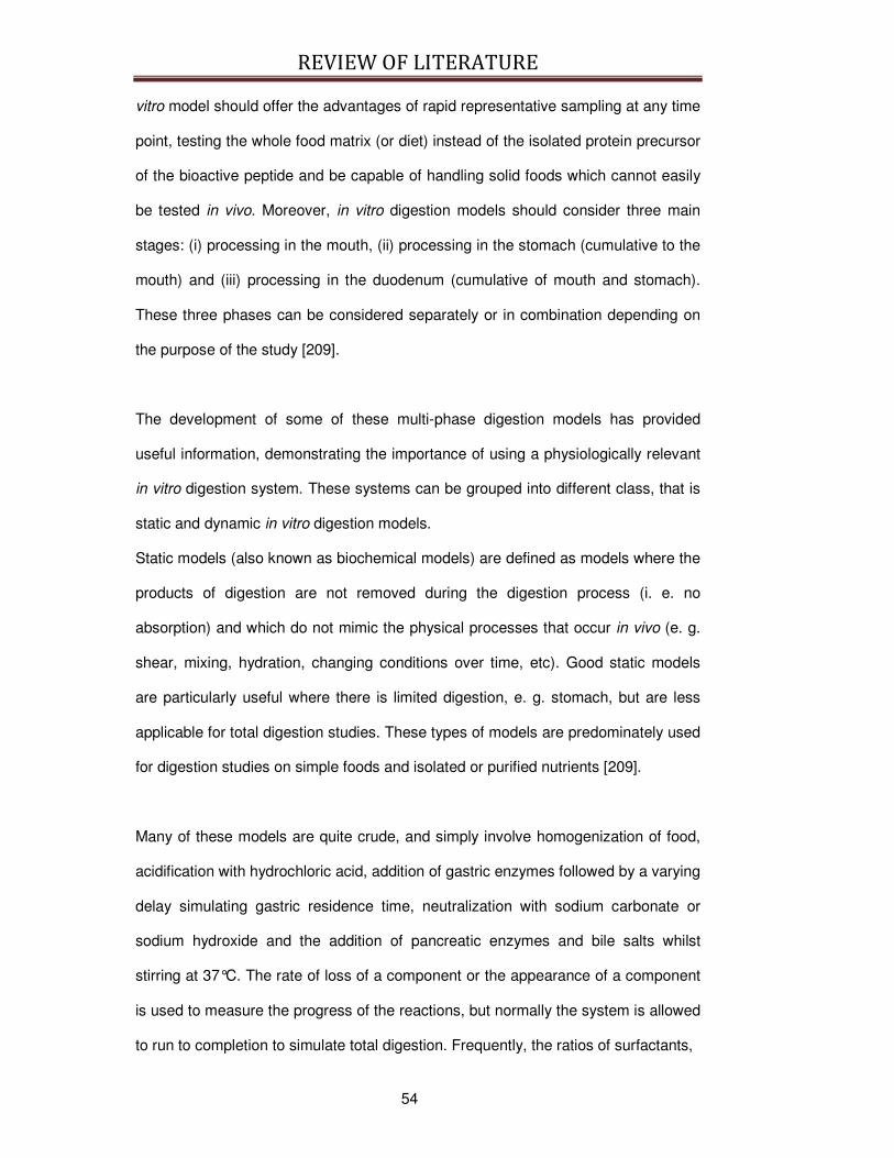
REVIEW OF LITERATURE
54
vitro model should offer the advantages of rapid representative sampling at any time
point, testing the whole food matrix (or diet) instead of the isolated protein precursor
of the bioactive peptide and be capable of handling solid foods which cannot easily
be tested in vivo. Moreover, in vitro digestion models should consider three main
stages: (i) processing in the mouth, (ii) processing in the stomach (cumulative to the
mouth) and (iii) processing in the duodenum (cumulative of mouth and stomach).
These three phases can be considered separately or in combination depending on
the purpose of the study [209].
The development of some of these multi-phase digestion models has provided
useful information, demonstrating the importance of using a physiologically relevant
in vitro digestion system. These systems can be grouped into different class, that is
static and dynamic in vitro digestion models.
Static models (also known as biochemical models) are defined as models where the
products of digestion are not removed during the digestion process (i. e. no
absorption) and which do not mimic the physical processes that occur in vivo (e. g.
shear, mixing, hydration, changing conditions over time, etc). Good static models
are particularly useful where there is limited digestion, e. g. stomach, but are less
applicable for total digestion studies. These types of models are predominately used
for digestion studies on simple foods and isolated or purified nutrients [209].
Many of these models are quite crude, and simply involve homogenization of food,
acidification with hydrochloric acid, addition of gastric enzymes followed by a varying
delay simulating gastric residence time, neutralization with sodium carbonate or
sodium hydroxide and the addition of pancreatic enzymes and bile salts whilst
stirring at 37°C. The rate of loss of a component or the appearance of a component
is used to measure the progress of the reactions, but normally the system is allowed
to run to completion to simulate total digestion. Frequently, the ratios of surfactants,
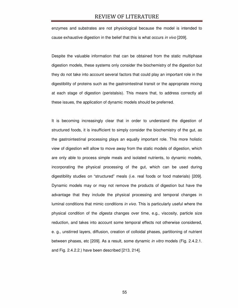
REVIEW OF LITERATURE
55
enzymes and substrates are not physiological because the model is intended to
cause exhaustive digestion in the belief that this is what occurs in vivo [209].
Despite the valuable information that can be obtained from the static multiphase
digestion models, these systems only consider the biochemistry of the digestion but
they do not take into account several factors that could play an important role in the
digestibility of proteins such as the gastrointestinal transit or the appropriate mixing
at each stage of digestion (peristalsis). This means that, to address correctly all
these issues, the application of dynamic models should be preferred.
It is becoming increasingly clear that in order to understand the digestion of
structured foods, it is insufficient to simply consider the biochemistry of the gut, as
the gastrointestinal processing plays an equally important role. This more holistic
view of digestion will allow to move away from the static models of digestion, which
are only able to process simple meals and isolated nutrients, to dynamic models,
incorporating the physical processing of the gut, which can be used during
digestibility studies on “structured” meals (i.e. real foods or food materials) [209].
Dynamic models may or may not remove the products of digestion but have the
advantage that they include the physical processing and temporal changes in
luminal conditions that mimic conditions in vivo. This is particularly useful where the
physical condition of the digesta changes over time, e.g., viscosity, particle size
reduction, and takes into account some temporal effects not otherwise considered,
e. g., unstirred layers, diffusion, creation of colloidal phases, partitioning of nutrient
between phases, etc [209]. As a result, some dynamic in vitro models (Fig. 2.4.2.1.
and Fig. 2.4.2.2.) have been described [213, 214].
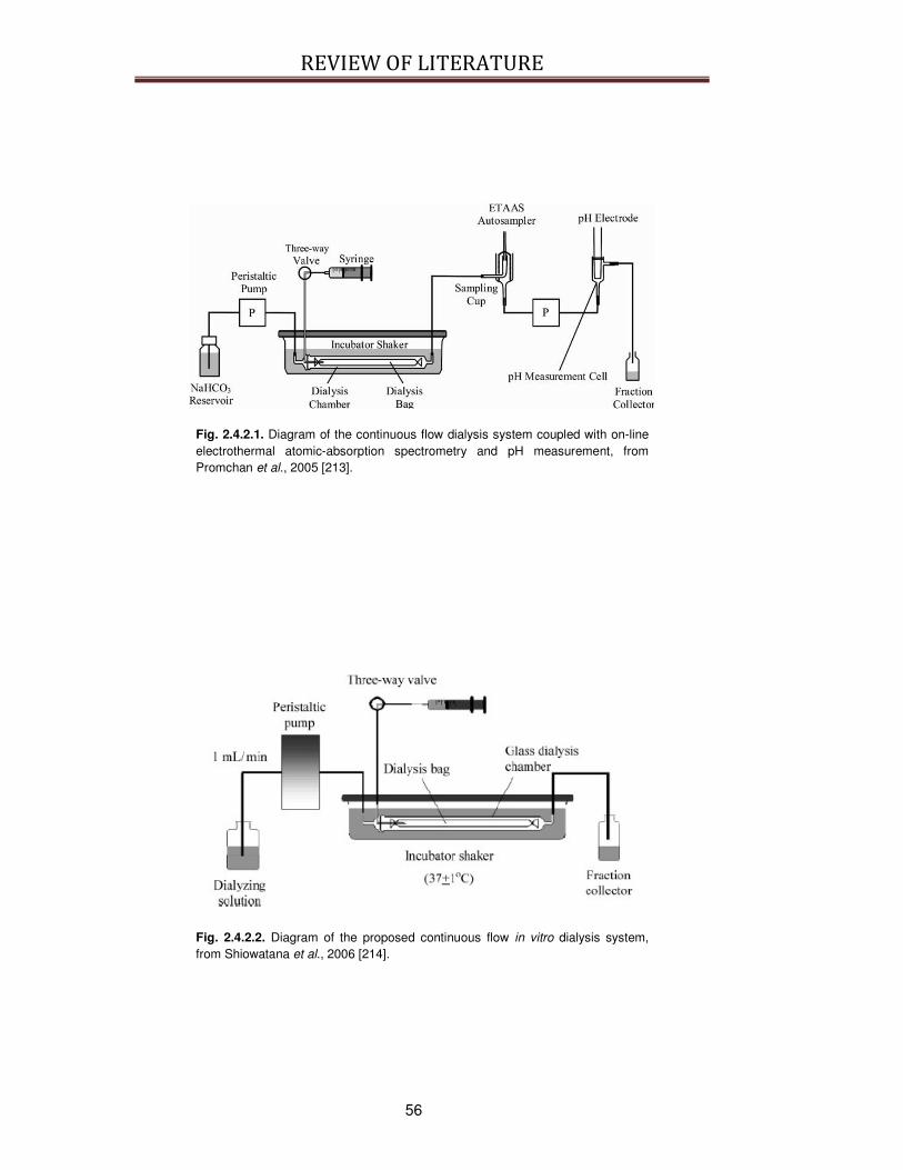
REVIEW OF LITERATURE
56
Fig. 2.4.2.1. Diagram of the continuous flow dialysis system coupled with on-line electrothermal atomic-absorption spectrometry and pH measurement, from Promchan et al., 2005 [213].
Fig. 2.4.2.2. Diagram of the proposed continuous flow in vitro dialysis system, from Shiowatana et al., 2006 [214].

REVIEW OF LITERATURE
57
An intermediated model for the evaluation of the release of ACE-inhibitory activity
during the digestion has been realized by Vermeirssen and colleagues [198]. This is
a semi-continuous model based on the batch physiological digestion (Fig. 2.4.2.3.).
In this reactor, the influence of temperature and incubation time in the stomach and
small intestine phase on the formation of ACE-inhibitory activity and the degree of
proteolysis is investigated.
Fig. 2.4.2.3. Experimental setup for the semi-continuous digestion, from Vermeirssen et al., 2003 [198].
More recently, Bastianelli and colleagues [215] explored the mathematical
modelization of the nutrient digestion in pig. This four-compartments model (see Fig.
2.4.2.4.) permitted to integrate various factors that normally affect food digestion.
This approach could be applied to evaluate the digestion of bioactive peptides,
because the porcine system has been shown to be a valid approximation of the
analogous systems in humans and has been used extensively to model human
digestion [216-218].
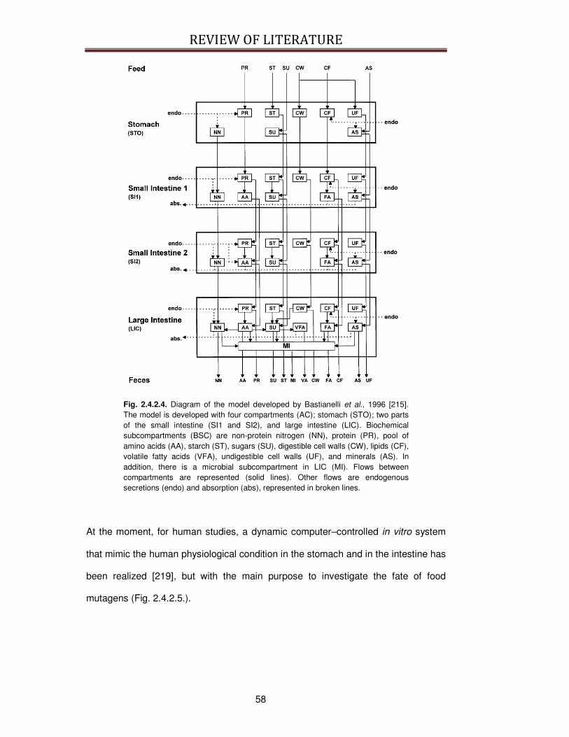
REVIEW OF LITERATURE
58
Fig. 2.4.2.4. Diagram of the model developed by Bastianelli et al., 1996 [215]. The model is developed with four compartments (AC); stomach (STO); two parts of the small intestine (SI1 and SI2), and large intestine (LIC). Biochemical subcompartments (BSC) are non-protein nitrogen (NN), protein (PR), pool of amino acids (AA), starch (ST), sugars (SU), digestible cell walls (CW), lipids (CF), volatile fatty acids (VFA), undigestible cell walls (UF), and minerals (AS). In addition, there is a microbial subcompartment in LIC (MI). Flows between compartments are represented (solid lines). Other flows are endogenous secretions (endo) and absorption (abs), represented in broken lines.
At the moment, for human studies, a dynamic computer–controlled in vitro system
that mimic the human physiological condition in the stomach and in the intestine has
been realized [219], but with the main purpose to investigate the fate of food
mutagens (Fig. 2.4.2.5.).

REVIEW OF LITERATURE
59
Fig. 2.4.2.5. Schematic diagram of the dynamic in vitro model of the stomach and the small intestine (TIM): (A) gastric compartment; (B) duodenal compartment; (C) jejunal compartment; (D) ileal compartment; (E) glass jacket; (F) flexible wall; (G) rotary pump; (H) pyloric valve; (I) pH electrodes; (J) secretion pump; (K) pre-filter; (L) hollow fiber membrane; (M) dialysis system; (N) ileal delivery valve; 1.1 detail of the hollow fiber membrane system, from Krul et al., 2000 [219].
An innovative approach to predict the uptake of iron in humans has been developed
by Glahn and colleagues [220]. This in vitro model (Fig. 2.4.2.6.) combines Caco-2
cell line in conjunction with in vitro digestion techniques and develops a model
whereby foods undergo simulated peptic digestion followed by intestinal digestion in
the presence of Caco-2 cell monolayers. The conditions of this model have been
designed to simulate the gastrointestinal environment while still maintaining a rapid
and inexpensive system. This model system is unique among applications of Caco-2
cells and in vitro digestion techniques in that it allows uptake to occur
simultaneously with food digestion under pH conditions similar to those found along
the absorptive surface of the intestinal mucosa. Furthermore, the addition of the
human-derived component, i.e., Caco-2 cells, transforms this model system into a
tool capable of conducting experiments that might not be feasible or practical to
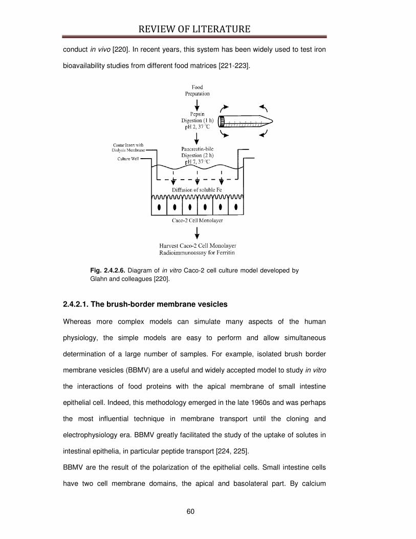
REVIEW OF LITERATURE
60
conduct in vivo [220]. In recent years, this system has been widely used to test iron
bioavailability studies from different food matrices [221-223].
Fig. 2.4.2.6. Diagram of in vitro Caco-2 cell culture model developed by Glahn and colleagues [220].
2.4.2.1. The brush-border membrane vesicles
Whereas more complex models can simulate many aspects of the human
physiology, the simple models are easy to perform and allow simultaneous
determination of a large number of samples. For example, isolated brush border
membrane vesicles (BBMV) are a useful and widely accepted model to study in vitro
the interactions of food proteins with the apical membrane of small intestine
epithelial cell. Indeed, this methodology emerged in the late 1960s and was perhaps
the most influential technique in membrane transport until the cloning and
electrophysiology era. BBMV greatly facilitated the study of the uptake of solutes in
intestinal epithelia, in particular peptide transport [224, 225].
BBMV are the result of the polarization of the epithelial cells. Small intestine cells
have two cell membrane domains, the apical and basolateral part. By calcium
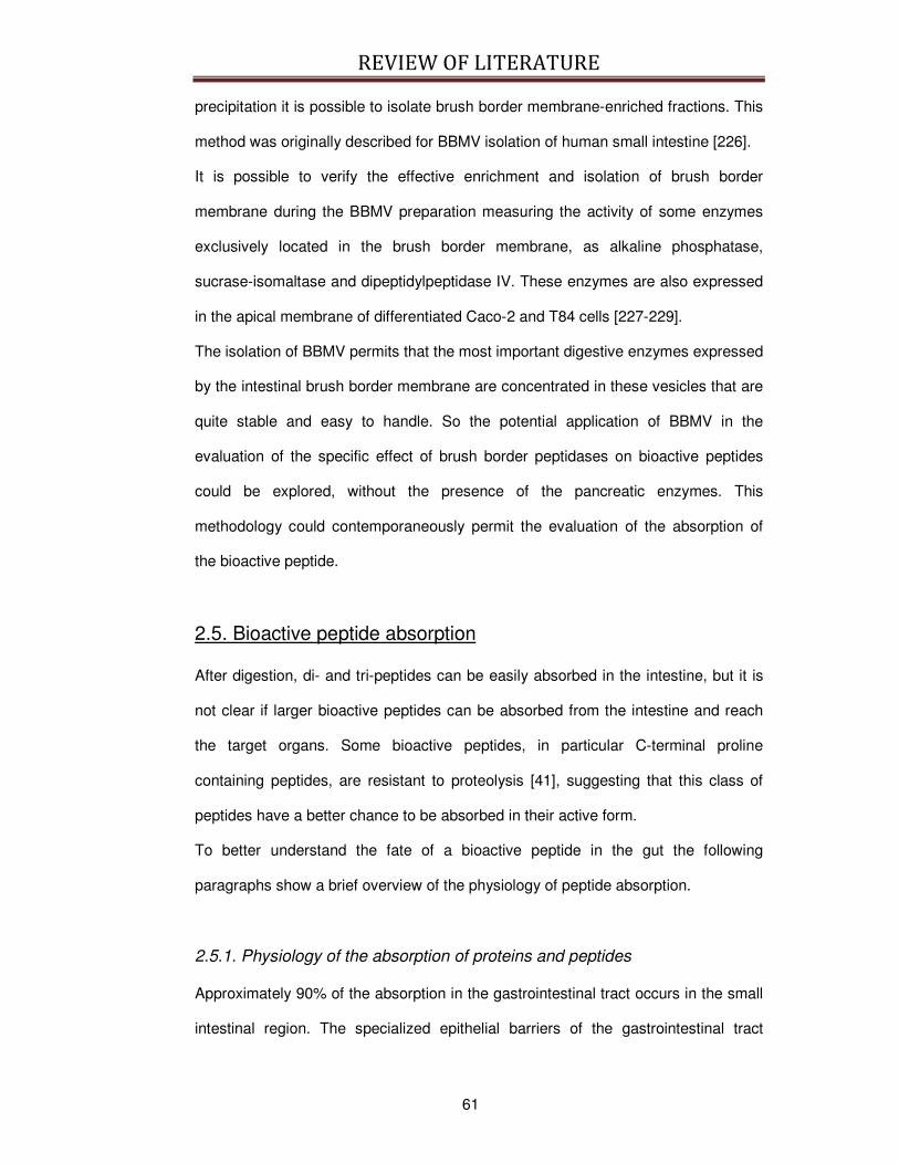
REVIEW OF LITERATURE
61
precipitation it is possible to isolate brush border membrane-enriched fractions. This
method was originally described for BBMV isolation of human small intestine [226].
It is possible to verify the effective enrichment and isolation of brush border
membrane during the BBMV preparation measuring the activity of some enzymes
exclusively located in the brush border membrane, as alkaline phosphatase,
sucrase-isomaltase and dipeptidylpeptidase IV. These enzymes are also expressed
in the apical membrane of differentiated Caco-2 and T84 cells [227-229].
The isolation of BBMV permits that the most important digestive enzymes expressed
by the intestinal brush border membrane are concentrated in these vesicles that are
quite stable and easy to handle. So the potential application of BBMV in the
evaluation of the specific effect of brush border peptidases on bioactive peptides
could be explored, without the presence of the pancreatic enzymes. This
methodology could contemporaneously permit the evaluation of the absorption of
the bioactive peptide.
2.5. Bioactive peptide absorption
After digestion, di- and tri-peptides can be easily absorbed in the intestine, but it is
not clear if larger bioactive peptides can be absorbed from the intestine and reach
the target organs. Some bioactive peptides, in particular C-terminal proline
containing peptides, are resistant to proteolysis [41], suggesting that this class of
peptides have a better chance to be absorbed in their active form.
To better understand the fate of a bioactive peptide in the gut the following
paragraphs show a brief overview of the physiology of peptide absorption.
2.5.1. Physiology of the absorption of proteins and peptides
Approximately 90% of the absorption in the gastrointestinal tract occurs in the small
intestinal region. The specialized epithelial barriers of the gastrointestinal tract
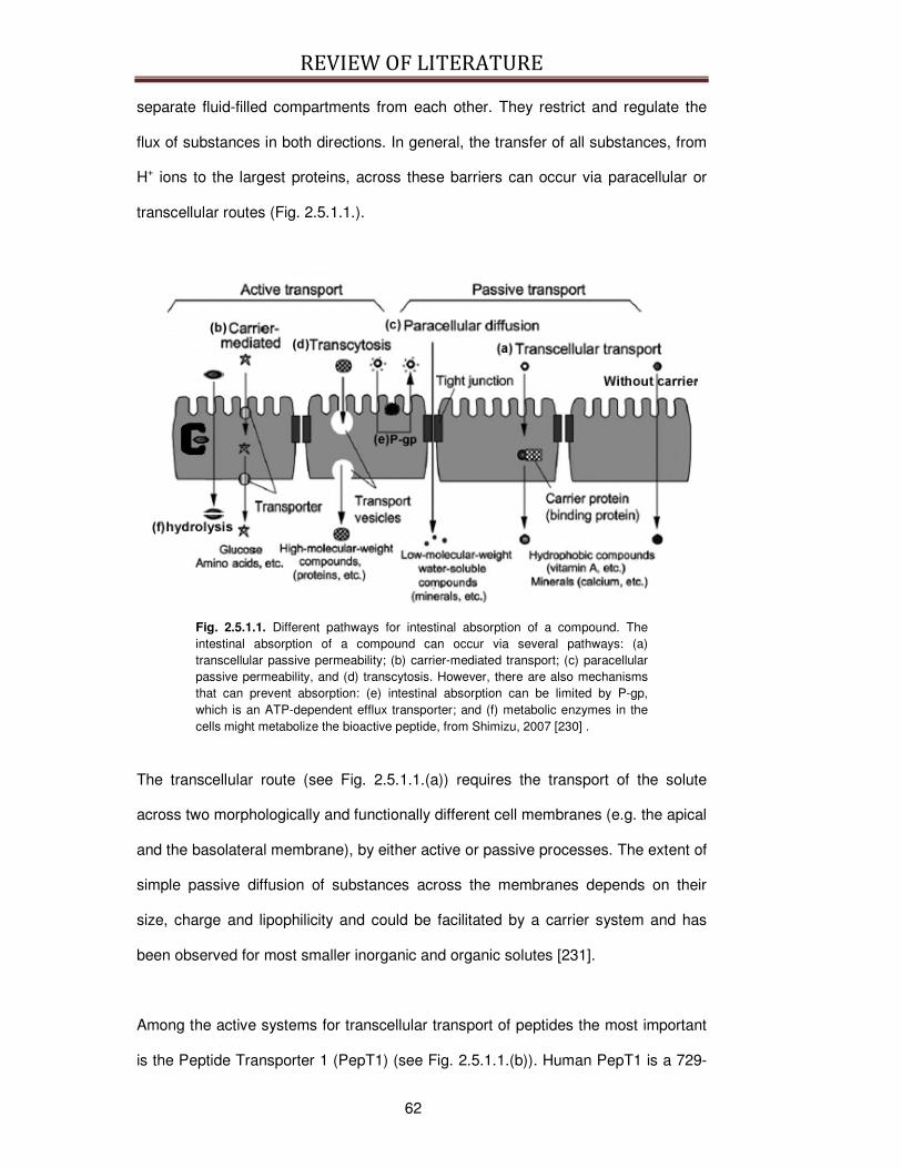
REVIEW OF LITERATURE
62
separate fluid-filled compartments from each other. They restrict and regulate the
flux of substances in both directions. In general, the transfer of all substances, from
H+ ions to the largest proteins, across these barriers can occur via paracellular or
transcellular routes (Fig. 2.5.1.1.).
Fig. 2.5.1.1. Different pathways for intestinal absorption of a compound. The intestinal absorption of a compound can occur via several pathways: (a) transcellular passive permeability; (b) carrier-mediated transport; (c) paracellular passive permeability, and (d) transcytosis. However, there are also mechanisms that can prevent absorption: (e) intestinal absorption can be limited by P-gp, which is an ATP-dependent efflux transporter; and (f) metabolic enzymes in the cells might metabolize the bioactive peptide, from Shimizu, 2007 [230] .
The transcellular route (see Fig. 2.5.1.1.(a)) requires the transport of the solute
across two morphologically and functionally different cell membranes (e.g. the apical
and the basolateral membrane), by either active or passive processes. The extent of
simple passive diffusion of substances across the membranes depends on their
size, charge and lipophilicity and could be facilitated by a carrier system and has
been observed for most smaller inorganic and organic solutes [231].
Among the active systems for transcellular transport of peptides the most important
is the Peptide Transporter 1 (PepT1) (see Fig. 2.5.1.1.(b)). Human PepT1 is a 729-
REVIEW OF LITERATURE
63
residues transmembrane protein complex with 12 transmembrane domains (see Fig.
2.5.1.2.) belonging to the proton-dependent oligopeptide transporter (POT) family.
This transporter is mostly a degradative way because cytosolic peptidases rapidly
hydrolyze most of the di- and tripeptides transported by PepT1. One of the most
commonly used and best known reference ways to study the peptide transport
mediated by of PepT1 is the substrate [14C]glycylsarcosine (Gly-Sar). Gly-Sar is
relatively stable against intra- and extracellular enzymatic hydrolysis and it acts as
competitive substrate of PepT1. Other labeled reference substrates quite often used
to study peptide transport by PepT1 are [3H]D-Phe-L-Ala, [3H]D-Phe-L-Gln and D-
Ala-L-Lys-Ne-7-amino-4-methylcoumarin-3-acetic acid [232].
Fig. 2.5.1.2. Membrane topology of human PepT1, from http://www.wzw.tum.de/nutrition/index.php?id=31.
The paracellular pathway (see Fig. 2.5.1.1.(c)) is very often restricted by tight
junctions (TJ), and the ability of substances to cross epithelia between the cells by
simple passive diffusion depends mainly on their size. TJ between intestinal
epithelial cells play an important role in the regulation of transport of organic and
inorganic compounds from the gut lumen towards blood circulation [233]. TJ
maintain the specificity of apical and basolateral domains and form a fence to
prevent mixing of apical and basolateral membrane components as well as an

REVIEW OF LITERATURE
64
occluding barrier between neighboring cells [234]. TJ membrane proteins interact
with scaffold proteins to connect them with various signal transduction and
transcriptional pathways involved in the regulation of TJ function [235]. Although the
permeability of TJ varies significantly within different epithelia, TJ are generally
reported to be impermeable to molecules with radii larger than 11-15 Å [236].
Paracellular passive diffusion process is applicable for a wide variety of low-
molecular weight compounds including peptides [186, 237, 238]. Moreover, it has to
be considered that TJ are affected by several extracellular stimuli, as nutrients, INFγ
and cytokines [239-242]. Thus the paracellular transport of small compounds, as
bioactive peptides, could be different in vivo than that predicted from the in vitro
approaches [243].
The potent mycotoxin cytochalasin D could be used to perturb the TJ. The
administration of cytochalasin D could be useful to study the mechanism of
absorption of those food-derived compounds whose transport could be mediated by
tight junction, as small peptides. Cytochalasin D acts as an inhibitor of actin
polymerization and it disrupts actin microfilaments. Several studies aiming to
elucidate the mechanism of absorption of bioactive peptides used Cytochalasin D to
increase the permeability of paracellular passive transport [201, 202, 244].
Large proteins or peptides that cannot be absorbed by PepT1 are translocated
across cell layers mainly by specialized transcytotic processes involving membrane
invagination and vesicle internalization (Fig. 2.5.1.1.(d)). Cellular internalization via
vesicles could be divided in fluid-phase endocytosis, that does not require any
interaction between the peptide and the apical membrane [245], and in receptor-
mediated absorptive endocytosis [246] that involves a binding with the plasma
membrane before being incorporated into endocytotic vesicles.
REVIEW OF LITERATURE
65
Once internalized inside the vesicles, the proteins or peptides are recycled back to
the plasma membrane or processed in the course of a multistep transport sequence
through various intracellular organelles, such as endosomes, prelysosomes and
lysosomes [247]. If the fusion with lysosomes does not completely disrupt the
endocytosed molecules, they could also be transported to the opposite cell surface
completing the transcytotic process [248].
Transcytosis could be studied by selective inhibitors of this pathway, as wortmannin.
Wortmannin was first discovered in 1957 in the broth of the fungi Penicillium
wortmannin Klocker [249] and it is a specific covalent inhibitor of phosphoinoside 3-
kinase and it is also involved in the inhibition of receptor-mediated endocytosis
[250]. Cardone and Mostov observed that wortmannin inhibits transcytosis in
epithelial cells, more specifically in those related to the mucosal immunity [251].
At present, numerous data demonstrate that dipeptides and tripeptides are
transported intact from the lumen into the enterocytes by the H+/peptide transporter
PepT1 [5, 6, 11]. Peptides resistant to cytosolic peptidases may be transported
intact across the basolateral membrane of intestinal cells by a peptide transport
system that has been characterized so far only at the functional level.
The mechanisms involved in the transfer of peptides across the intestinal
basolateral membrane to the blood side are still under debate. The investigation by
Dyer et al. [252] using rabbit enterocyte basolateral membrane vesicles was the first
to study basolateral peptide transport. This report described a system relatively
specific for small peptides that, just as PepT1 in the apical membrane, is stimulated
by an inwardly directed H+ gradient. Yet, the H+ gradient across the basolateral
membrane is expected to be very small. This might provide the basis for
transcellular movement of small peptides across the enterocyte despite the fact that
the peptide transport systems in both poles of the cell are H+ dependent [253].
Terada et al. [254] reported that uptake of [14C]Gly-Sar across the basolateral

REVIEW OF LITERATURE
66
membrane in Caco-2 cells cultured on filters was less sensitive to extracellular pH
than uptake across the apical membrane by PepT1. Importantly, the uptake did not
proceed against a concentration gradient. This result led to the conclusion that the
basolateral system is a facilitative peptide transporter whereas PepT1 is an active
transporter [254]. If few information is available on basolateral transport of small
peptides, still less is known on the basolateral transport of large peptides, that is
mainly mediated by an esocytotic vesicles.
In conclusion, the intestinal transport of peptides is not fully elucidated and many
questions remain open. For example, the mechanism by which the transport
systems for peptides are differentially sorted in the enterocyte to be inserted into the
brush border and/or the basolateral membrane is still controversial. Another serious
lack of knowledge exists about the number of carriers per cell and the substrate
turnover rates. The identity of the postulated basolateral peptide transporters
remains to be elucidated. The intestinal absorption of ACE inhibitors needs to be re-
evaluated. Furthermore, inter-individual differences in peptide transport should be
one of the priorities of future research in this area [231].
2.5.2. Physical and chemical characteristics of potentially absorbable
bioactive peptides
To exert physiological effects after oral ingestion, it is of crucial importance that milk-
derived bioactive peptides remain active during gastrointestinal digestion and
absorption and reach the circulation. The bioavailability of peptides depends on a
variety of structural and chemical properties, i.e. resistance to proteases, charge,
molecular weight, hydrogen bonding potential, hydrophobicity and the presence of
specific residues [192, 253, 255]. Indeed, proline- and hydroxyproline-containing
peptides are relatively resistant to degradation by digestive enzymes [41, 256, 257].

REVIEW OF LITERATURE
67
Furthermore, tripeptides containing the C-terminal proline–proline are reported to be
resistant to proline-specific peptidases [73] and have been shown to be stable under
simulated gastrointestinal digestion conditions [41]. As already explained in
paragraph 2.5.1., peptides consisting of two or three amino acids can be absorbed
intact from the intestinal lumen into the blood circulation via different mechanisms
for intestinal transport [54]. The presence of the milk-derived ACE-inhibitory peptide
IPP was recently demonstrated in measurable amounts in the circulation of
volunteers that consumed a drink enriched in IPP and VPP [6].
Other characteristics contribute to the resistance to hydrolysis. For example, when
isolated, some casein-derived peptides tend to be highly negatively charged and
phosphorylated, making them resistant to further proteolysis [258]. Thus, some of
the bioactive peptides could be absorbed across the intestinal mucosa to enter the
circulation or be retained in the lumen and pass into the colon. The latter is likely
based on evidence that ingested casein-derived phosphopeptides can be isolated
from rat feces [259].
2.5.2.1. The absorption of bioactive peptides derived from milk proteins
For some bioactive tripeptides the intestinal absorption has been already
demonstrated. For example, VPP was detected in the abdominal aorta of SHR 6
hours after its administration in sour milk, which strongly suggests that it is
transepithelially transported [260]; more recently the absorption was observed also
in humans [6]. Paracellular transport, through the intercellular junctions, was
suggested as the main mechanism, since the transport via the short-peptide carrier,
PepT1, led to a quick hydrolysis of the internalized peptide [186]. In the case of
larger sequences, the susceptibility to brush border peptidases is the primary factor
that decides the transport rate [244]. For example, the heptapeptide lactokinins
(ALPMHIR) was transported intact, although in concentrations too low to exert an
ACE-inhibitory activity, which suggests cleavage by aminopeptidases [261].

REVIEW OF LITERATURE
68
2.5.3. Absorption models
Early studies on peptide transport were performed in feeding or perfusion
experiments in vivo and in situ. Tissue, cell and membrane preparations, such as
the Ussing chamber technique (see Fig. 2.5.3.1.), the everted gut sac or ring
technique (see Fig. 2.5.3.2.), and brush-border membrane vesicles, have been used
for at least 50 years and are still being used today. During times, other methods
have been realized to study peptide absorption and the variety of the systems can
be grouped into three main groups: in vivo, ex vivo and in vitro. None of them is
completely exhaustive and so a combination of some of them is necessary to
understand if a peptide of interest can be absorbed.
Fig. 2.5.3.1. Representation of the Ussing chamber technique, from http://www.warneronline.com/product_info.cfm?name=Introduction to Ussing Chamber and System from Warner&id=1401.

REVIEW OF LITERATURE
69
Fig. 2.5.3.2. Representation of the everted gut sac technique.
The in vivo methods usually evaluated the concentration of the labeled bioactive
peptide of interest in the circulation and thus they permitted to understand if a
peptide crosses the intestinal barrier and if it distributes in blood in enough amount
to reach the target site, exhibiting its bioactivity. In vivo studies have been performed
in humans [11, 82, 104, 105] or animals [72, 78, 79, 97-103] with a GI system
supposed to be comparable to human GI system. However, only little information
can be acquired about the transport mechanism at molecular level in the intestinal
epithelium.
The ex vivo methods are a good compromise between the in vivo and the in vitro
systems because they take into consideration the intestinal tissue complexity and
organization, and some information on transport mechanism at molecular level can
be acquired. In fact, these methodologies evaluate the absorption of the molecule of
interest sampling a part of the intestinal tube. However, compared to the in vivo
systems, they give insufficient information on the fate and the stability of the
bioactive peptide, once absorbed and in the blood. In addition, they are not always
so easy to perform. The ex vivo methods include various techniques, briefly
explained here. For example, the everted gut sac (see Fig. 2.5.3.2.), that is a simple
and useful model first employed to study drug transport [262]. This methodology

REVIEW OF LITERATURE
70
consists of a freshly excised small intestine incubated in appropriate tissue medium
and the content of the molecule of interest is tested in the intestinal sac. The everted
gut sac has been used to study the uptake of lipid vesicles [263], proteins and
macromolecules with oral drug delivery potential. It is useful because it provides
quantitative information on the uptake and absorption of the tested compound [264].
Another ex vivo technique is the in situ perfusion system that monitor the
disappearance from the lumen of the gut segment and the measurement of plasma
concentrations of the molecule following perfusion [265]. Although disappearance
from the lumen in many cases may provide an adequate estimation of absorption,
caution in interpretation of results from studies with this technique is warranted since
an overestimation of absorption due to biotransformation, binding, and/or partitioning
can occur [266].
Isolated intestinal tissues have been employed to determine uptake of oligopeptides
across the apical membrane and transepithelial transport [267-270]. The major
issues associated with the use of isolated tissues are the life-span of preparations
and the metabolic activity of enterocytes, which often precludes transepithelial
transport studies due to intracellular hydrolysis [270]. In this class of ex vivo
absorption models The Ussing chamber technique (see Fig. 2.5.3.1.) has been
applied to the study of transepithelial transport mechanisms of various compounds.
Tissue preparations in Ussing chambers have been demonstrated to be viable (from
both electrical measurements and transepithelial flux studies) and to maintain their
integrity (based on electrical measurements and flux studies with passive
permeability markers) for several hours in vitro. So tissue studies provide a
convenient and rapid method for assessing mechanisms involved in transepithelial
transport and segmental differences in these transport processes [264].
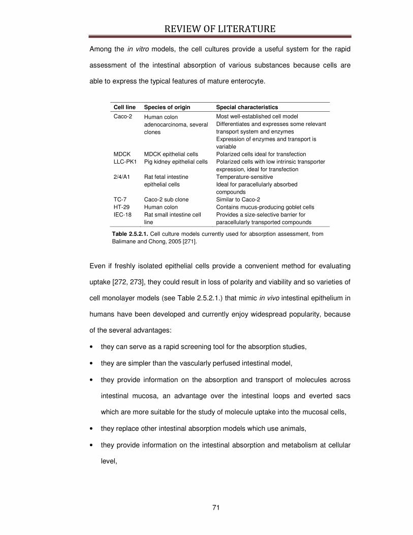
REVIEW OF LITERATURE
71
Among the in vitro models, the cell cultures provide a useful system for the rapid
assessment of the intestinal absorption of various substances because cells are
able to express the typical features of mature enterocyte.
Cell line Species of origin Special characteristics
Caco-2 Human colon adenocarcinoma, several clones
Most well-established cell model Differentiates and expresses some relevant transport system and enzymes Expression of enzymes and transport is variable
MDCK MDCK epithelial cells Polarized cells ideal for transfection LLC-PK1 Pig kidney epithelial cells Polarized cells with low intrinsic transporter
expression, ideal for transfection 2/4/A1 Rat fetal intestine
epithelial cells Temperature-sensitive Ideal for paracellularly absorbed compounds
TC-7 Caco-2 sub clone Similar to Caco-2 HT-29 Human colon Contains mucus-producing goblet cells IEC-18 Rat small intestine cell
line Provides a size-selective barrier for paracellularly transported compounds
Table 2.5.2.1. Cell culture models currently used for absorption assessment, from Balimane and Chong, 2005 [271].
Even if freshly isolated epithelial cells provide a convenient method for evaluating
uptake [272, 273], they could result in loss of polarity and viability and so varieties of
cell monolayer models (see Table 2.5.2.1.) that mimic in vivo intestinal epithelium in
humans have been developed and currently enjoy widespread popularity, because
of the several advantages:
• they can serve as a rapid screening tool for the absorption studies,
• they are simpler than the vascularly perfused intestinal model,
• they provide information on the absorption and transport of molecules across
intestinal mucosa, an advantage over the intestinal loops and everted sacs
which are more suitable for the study of molecule uptake into the mucosal cells,
• they replace other intestinal absorption models which use animals,
• they provide information on the intestinal absorption and metabolism at cellular
level,
REVIEW OF LITERATURE
72
• they provide information on the possible mucosal toxicity caused by the
substance of interest,
• most of them do not need interspecies correlation because of human origin.
Thus, for the peptides whose in vitro permeability reflects in vivo permeability, cell
culture can be effectively used a first evaluation step.
Among the various cell lines, colon carcinoma cell lines grow relatively rapidly into
confluent monolayers and exhibit a differentiated absorptive phenotype under
certain culture conditions. Therefore, they have been used as a tool for studying
enterocytic differentiation and function, including cell structure [274], brush border
morphogenesis [275], synthesis and localization of brush border enzymes [229,
276], electrolyte transport [277] and amino acid/protein uptake [227, 278]. The most
employed colon carcinoma cell lines are mostly three; HT-29, which is
undifferentiated when grown under standard culture conditions and expresses
enterocytic differentiation only after deprivation of glucose from the culture media
[279] or addition of certain inducers [280], then the cell lines Caco-2 and T84 that
spontaneously differentiate at confluence and show features of small intestinal
enterocytes [274, 277, 281, 282].
Despite the advantages, a clear limitation of these systems is that intestinal
segmental differences in transport cannot be discerned. In addition, the cell model
composed of solely absorptive cells may be a oversimplified system, because the
intestinal epithelium is a conglomerate of absorptive enterocytes and other cells
such as mucous-secreting cells (the second most frequent cell type), endocrine
cells, and M cells [283].
Another tool to evaluate in vitro the absorption of different molecules is constituted
by the vesicles isolated from brush border membrane. At present, results from

REVIEW OF LITERATURE
73
membrane vesicle studies have not been consistent because of the extreme
variability during vesicles preparation and the inability to monitor the internal milieu
prior to experiment. For example, this experimental issue was observed for the pH
dependent overshoot phenomena of oligopeptide transport that has been
demonstrated in some but not all studies [284]. The “leakiness” of vesicle
preparations or lack of appropriate conditions at the time of the experiments may
also account in part for the differences observed.
2.5.3.1. The Caco-2 cell line model
An accepted model system for the enterocyte of the human small intestine is the
Caco-2 cell line (Fig. 2.5.3.1.1.), as - with ongoing differentiation - this colon
carcinoma cells exhibit morphological and functional similarities to non-malignant
human enterocytes (as cell polarization, expression of brush border enzymes,
formation of tight junctions (TJ), the microvillous structure, the carrier-mediated
transport system for di- and tri-peptides and amino acids PepT1) [285].
Fig. 2.5.3.1.1. Caco-2 cell monolayer, from www.fi.cnr.it/r&f/n4/images/spadoni.jpg
The TJ which regulates the paracellular transport of the cell monolayer has also
been expressed in Caco-2 cell monolayers cultured on a semipermeable filter. In
addition, transcytotic activity has also been observed in Caco-2 cells.
TJ function of Caco-2 cells can be determined and monitored by the measurement
of TransEpithelial Electrical Resistance (TEER) using a two compartment cell
culture system, separating the upper (apical) part of the epithelium from a

REVIEW OF LITERATURE
74
basolateral compartment mimicking the cellular sites facing blood circulation [195].
One main advantage of the TEER assay is that it is non-destructive and changes in
TEER and consequently TJ permeability can be monitored over a long period of
time [286]. Fig. 2.5.2.1.2. describes the measurement in Caco-2 cell.
Fig. 2.5.2.1.2. Transepithelial electrical resistance principle of measurement, from Hartmann et al, 2007 [195].
Integrity of cultured monolayers is also detectable by carrying out transport studies
using water-soluble reference compounds that can be absorbed by TJ channels
between the cells (e.g. radio-labeled mannitol, phenol red, Lucifer Yellow, or
flourescein; Mr: 182, 354, 57, 332 Da, respectively). Quantification is then made by
detecting the reference substance in the basolateral compartment [287].
Pure Caco-2 cell system shows some limitations, as the potential overexpression of
the P-glycoprotein (P-gp), which may lead to higher excretion rates of the tested
molecules (i. e. the bioactive peptide of interest) and consequently lower
permeabilities in the absorptive direction [288]. In addition, because of the absence
of a prominent mucus layer on the surface of Caco-2 cell monolayers produced in
vivo by goblet cells, the apical pH will mainly be determined by the culture medium
[289], normally fixed at pH 7.4.
REVIEW OF LITERATURE
75
A better prediction of the absorption could be gained, if the apical pH is 5.5–6.5 and
this can be achieved without compromising the integrity of Caco-2 cell monolayers,
as demonstrated by Palm and colleagues [289] and Yamashita and colleagues
[290]. The change in pH has been evaluated in permeability studies for passively
permeated drugs [291] and the authors found that Caco-2 cells better mimicked the
in vivo conditions and gave more reliable information about the absorption of drugs
across the enterocytic membrane.
Finally, it is well known [292] that permeabilities of compounds that are transported
via carrier-mediated absorption are lower in the Caco-2 cell system as compared to
the human small intestine, probably also reflecting the colonic origin of this cell line.
In recent years several mucus-producing goblet cell sublines have been established
from human intestinal HT29 cells, as HT29-MTX [293-295], a cell population that
consists exclusively of differentiated, gastric-like mucus secreting, goblet-type cells
that retain their differentiated phenotype after reversion to a methotrexate (MTX)-
free medium and they also can be grown in monolayers.

EXPERIMENT 1
77
EXPERIMENT 1: Fermented milk from Enterococcus faecalis TH563 or Lactobacillus delbrueckii bulgaricus LA2 manifests different degrees of ACE-inhibitory and immunomodulatory activities
3.1. Introduction
There is evidence that several food or food ingredients provide a benefit beyond the
nutrients they contain. These substances are defined as functional food and their
putative biological effects have been extensively studied. To date, antihypertensive
and immunomodulatory bioactivities are frequently exploited in the production of
foodstuffs formulated to provide putative health benefits [9, 296].
The bioactive properties of fermented milks are often correlated to the generation of
specific peptides from milk proteins. The bioactive peptides are inactive when
encripted in the sequence of the precursor proteins but can be released by
enzymatic proteolysis during intestinal digestion or food processing [7]. Interestingly,
Angiotensin-I Converting Enzyme (ACE) inhibitory and immunomodulatory
properties seem to be associated, possibly because both are correlated to the
presence of short chain peptides [65].
So far, lactic acid bacteria have been preferred to others microorganisms to produce
fermented milks rich in ACE-inhibitory activity [40], in particular Lactobacillus
helveticus (L. helveticus) [12, 86], Lactobacillus delbrueckii subsp. bulgaricus (L.
delb. bulgaricus) and Lactococcus lactis subsp. cremoris (L. lactis cremoris) [29].
Moreover, some bacterial strains, mostly lactic acid bacteria, release components
during fermentation that possess immunomodulatory activity [4, 7]. Lactic acid
bacteria fermentation products potentiate the cell-mediated immune response by
increasing the proliferative response of lymphocytes to concanavalin A (conA), a
known activator of lymphocyte proliferation [297]. In addition, some findings suggest
that milk fermented by Lactobacillus strains can modulate the immune response
EXPERIMENT 1
78
against breast cancer cells in mice [4] and improve innate-defense capacity in
human [7].
However, species other than those belonging to Lactobacillus genus are often
isolated from dairy products, which may possess interesting properties [41, 188]
The aim of our study was to measure the ACE-inhibitory and immunomodulatory
bioactivities in milk fermented with Enterococcus faecalis TH563 and compare them
to those generated by L. delb. bulgaricus LA2. These strains belong to a panel of 14
bacterial strains (7 L. delb. lactis, 2 L. delb. bulgaricus, 1 L. helveticus, 2 L.
paracasei and 2 E. faecalis) representing species that are frequently isolated from
traditional dairy products of North Eastern Italy and showing different degrees of
proteolytic activity. The focus of the present study in E. faecalis was because it is an
enterococcal species frequently found in dairy products, traditional cheeses in
particular, where it may play an important role in determining chese taste and
texture [298, 299]. Altough E. faecalis is reported to generate fermented milk with
ACE-inhibitory activity [79, 112, 300, 301] few information about its ability to
generate immunomodulatory activity is available. On the contrary, L. delb.
bulgaricus is commonly used as the starter culture for the production of yogurt and
fermented milks, and it may represent a fairly well known control.
3.2. Materials and Methods
3.2.1. Chemicals and Reagents
Hank's balanced salt solution (HBSS) was purchased from Lonza, Switzerland.
Gibco-Invitrogen (United Kindom) supplied L-Glutamine (L-Glu). Lymphoprep was
purchased from Axis-Shield, Norway. Sigma–Aldrich (Italy) supplied Angiotensin-
converting enzyme (ACE), concanavalin A (conA), ethyl acetate, hippuryl-histidyl-
leucine (HHL), HCl, 3-(4,5-dimethyl-2-thiazolyl)-2,5-diphenyl-2H-tetrazolium bromide
(MTT) powder, new-born calf serum (NCS), NaOH, penicillin-streptomycin solution,

EXPERIMENT 1
79
RPMI-1640 medium, and Triton X100. Na borate buffer and NaCl were obtained
from Carlo Erba, Italy. MRS broth and sterilized skim milk were supplied by Biolife,
Italy and M17 broth by Difco Laboratories, Michigan, USA.
3.2.2. Bacteria culture
E. faecalis TH563 and L. delb. bulgaricus LA2 were evaluated for their proteolytic
activity as described by Hull [302] and in accordance with International Dairy
Federation (IDF) standard 149A (1997) [303].
Lactobacilli were propagated in MRS broth for 24 h at 44 °C, while enterococci were
propagated in M17 broth for 24 h at 37 °C. Revitalized microorganisms were used to
inoculate (1%, v/v) 10 mL of sterilised skim milk, which was incubated for 24 h at
44°C (lactobacilli) and 37 °C (enterococci). One mL of these milk pre-cultures was
used to inoculate 100 mL of skim milk. Incubation was carried out under sterile
conditions at 44 °C (lactobacilli) and 37 °C (enterococci). Fermented milk was
produced with skim milk under sterile conditions in order to avoid the presence of
enzyme interference by contaminating microorganisms.
3.2.3. Separation of the peptide fraction
Fermented milk samples were centrifuged at 20000 × g for 15 min at 15 °C (J2-21
Beckman Coulter centrifuge, JA 20 rotor, Fullerton, California, USA) to remove
bacteria debris. The supernatant was filtered with Amicon Centricon Ultra15
(molecular weight cut-off 5000 Da; Millipore, Billerica, Massachusetts, USA) by
centrifugation at 3200 × g for 40 min at 15° C. The fraction with molecular weight
lower than 5000 Da (5000 Da fraction) was stored at -20 °C and used for further
analyses. The concentration of peptides in the 5000 Da fractions was
spectrophotometrically determined by the method of Layne [304].

EXPERIMENT 1
80
3.2.4. ACE-inhibitory activity
The ACE-inhibitory activity of the 5000 Da fractions was measured by the method of
Cushman and Cheung [91], as modified by Nakamura and colleagues [86]. An
Ultrospec 3000 spectrophotometer (Amersham Pharmacia Biotech, New Jersey,
USA) was used to measure the optical density of each 5000 Da fraction.
Each test was performed in triplicates and the measured absorbance was used for
the calculation of the percentage of ACE inhibition (% ACE-I) as follows:
% ACE-I = 100 · (B − A) ÷ (B − C),
where A is the optical density of the samples in the presence of ACE, B is the optical
density of the total activity, and C is the optical density of the blank. Data underwent
analysis of variance and differences between mean values were analysed by the
test of Duncan (SPSS Inc., Chicago, Illinois, USA).
3.2.5. Bovine peripheral blood lymphocytes proliferation
Ten mL of 5000 Da fraction of fermented milk by E. faecalis TH563 and 30 mL of
5000 Da fraction of fermented milk by L. delb. bulgaricus LA2 were dried under
vacuum and the obtained powders were dissolved in 5 mL of complete medium
prepared as follow: RPMI-1640 medium containing 10 % of NCS, 2 mmol/L of L-Glu,
100 µg/mL of streptomycin and of 100 U/mL of penicillin. The concentration of
peptides in the 5000 Da fraction for the proliferation test was determined
spectrophotometrically as described by Layne [304]. The 5000 Da fractions were
sterilized by filtration (0.22 µm filters) and stored at -20 °C until use.
Bovine peripheral blood lymphocytes (BPBL) were isolated from whole heparin-
anticoagulated blood of nine non-pregnant, non-lactating dairy cows without clinical
symptoms by density gradient centrifugation using the Lymphoprep reagent. Cells
were suspended in completed medium in the presence of 2 µg/mL of conA as
mitogen and were incubated at 37 °C in 5% CO2. After 24 h of differentiation, non
EXPERIMENT 1
81
adherent BPBL were separated from adherent leukocytes and tested for viability
with Trypan blue staining. Viable BPBL were adjusted at density of 3·106 cells/mL in
complete medium and incubated for 48 h in a 96-well microplate (100 µL cell
suspension per well) with or without conA (2 µg/mL, positive control) and in
presence of increasing concentrations (from 0 µg/mL to 100 µg/mL) of each
fermented milk. At the end of the incubation period, proliferation test was assessed
by 3-(4,5-Dimethyl-2-thiazolyl)-2,5-diphenyl-2H-tetrazolium bromide (MTT)
proliferation test, following the manufacturer’s instructions. Briefly, MTT powder was
dissolved in Hanks’ balanced salt solution (5 mg/mL), added to the cells (15 µL per
well) and incubated for 3 h to allow the reductases of living cells to convert the MTT
into the insoluble formazan. The formazan was then eluted with 10% (v/v) Triton
X100 and the absorbance was measured at a wavelength of 570 nm with
background subtraction at 630 nm using a microplate reader (Spectra Count,
Packard Bioscience).
Each cell proliferation test was performed in triplicates. The results were expressed
as the percentage of the optical density observed in the conA-treated BPBL (%
conA). Relative variations of cellular proliferation produced by each fermented milk
were analysed using a Generalised Linear Model (GLM, SPSS Inc.). Differences
between mean values were analysed by the test of Dunnett (SPSS Inc.).
3.3. Results
E. faecalis TH563 and L. delb. bulgaricus LA2 showed a proteolytic activity of 0.292
and 0.100 mg of tyrosine/mL, respectively. The peptide concentration in the 5000
Da fraction was greater in milk fermented by E. faecalis TH563 than in milk
fermented by L. delb. bulgaricus LA2 (14.78 mg/mL and 4.89 mg/mL, respectively).
Milk fermented by E. faecalis TH563 showed a significantly (P < 0.05) higher ACE-
inhibitory activity (69.43 % ± 3.12) than L. delb. bulgaricus LA2 (60.86 % ± 1.01).
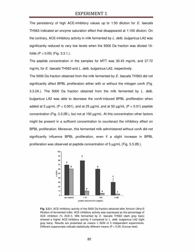
EXPERIMENT 1
82
The persistency of high ACE-inhibitory values up to 1:50 dilution for E. faecalis
TH563 indicated an enzyme saturation effect that disappeared at 1:100 dilution. On
the contrary, ACE-inhibitory activity in milk fermented by L. delb. bulgaricus LA2 was
significantly reduced to very low levels when the 5000 Da fraction was diluted 10-
folds (P < 0.05) (Fig. 3.3.1.).
The peptide concentration in the samples for MTT was 30.43 mg/mL and 37.72
mg/mL for E. faecalis TH563 and L. delb. bulgaricus LA2, respectively.
The 5000 Da fraction obtained from the milk fermented by E. faecalis TH563 did not
significantly affect BPBL proliferation either with or without the mitogen conA (Fig.
3.3.2A.). The 5000 Da fraction obtained from the milk fermented by L. delb.
bulgaricus LA2 was able to decrease the conA-induced BPBL proliferation when
added at 5 µg/mL (P < 0.001), and at 25 µg/mL and at 50 µg/mL (P < 0.01) peptide
concentration (Fig. 3.3.2B.), but not at 100 µg/mL. At this concentration other factors
might be present in a sufficent concentration to counteract the inhibitory effect on
BPBL proliferation. Moreover, this fermented milk administered without conA did not
significantly influence BPBL proliferation, even if a slight increase in BPBL
proliferation was observed at peptide concentration of 5 µg/mL (Fig. 3.3.2B.).
Fig. 3.3.1. ACE-inhibitory activity of the 5000 Da fraction obtained after Amicon Ultra15 filtration of fermented milks. ACE-inhibitory activity was expressed as the percentage of ACE inhibition (% ACE-I). Milk fermented by E. faecalis TH563 (dark grey bars) showed a higher ACE-inhibitory activity if compared to L. delb. bulgaricus LA2 (light grey bars). Results are presented as means ± SEM of 3 independent experiments. Different superscripts indicate statistically different means (P < 0.05; Duncan test).
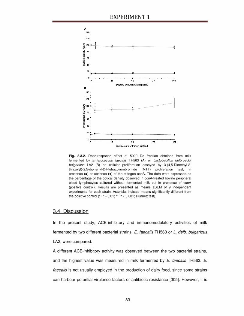
EXPERIMENT 1
83
Fig. 3.3.2. Dose-response effect of 5000 Da fraction obtained from milk fermented by Enterococcus faecalis TH563 (A) or Lactobacillus delbrueckii
bulgaricus LA2 (B) on cellular proliferation assayed by 3-(4,5-Dimethyl-2-thiazolyl)-2,5-diphenyl-2H-tetrazoliumbromide (MTT) proliferation test, in presence () or absence () of the mitogen conA. The data were expressed as the percentage of the optical density observed in conA-treated bovine peripheral blood lymphocytes cultured without fermented milk but in presence of conA (positive control). Results are presented as means ±SEM of 9 independent experiments for each strain. Asterisks indicate means significantly different from the positive control (* P > 0.01; ** P < 0.001; Dunnett test).
3.4. Discussion
In the present study, ACE-inhibitory and immunomodulatory activities of milk
fermented by two different bacterial strains, E. faecalis TH563 or L. delb. bulgaricus
LA2, were compared.
A different ACE-inhibitory activity was observed between the two bacterial strains,
and the highest value was measured in milk fermented by E. faecalis TH563. E.
faecalis is not usually employed in the production of dairy food, since some strains
can harbour potential virulence factors or antibiotic resistance [305]. However, it is

EXPERIMENT 1
84
frequently found in traditional cheeses, where it plays an important role in
determining cheese taste and texture [298]. Even if strains of E. faecalis have been
reported to possess high proteolytic activity [299], the ability to produce fermented
milks with ACE-inhibitory activity has been scarcerly documented [112, 301]. In the
present experiment, ACE inhibitory activity seemed to be positively related to the
proteolytic activity of the strain of interest. In fact, E. faecalis TH563 showed the
highest proteolytic activity and the highest peptide concentration in the 5000 Da
fraction, suggesting potentially greater ability to produce small peptides, which are
the main responsible of ACE inhibitory activity [306] .
In this experiment, ACE-inhibitory and immunomodulatory activities were not
associated, differently from the assumption of Narva and colleagues [65]. In fact, E.
faecalis TH563 did not alter BPBL proliferation, while L. delb. bulgaricus LA2 slightly
but significantly inhibited BPBL proliferation at low concentrations in presence of
conA. Both bacterial strains could not affect proliferation of BPBL keep in culture
without conA. This result supports the hypothesis of Fujiwara and colleagues [307]
suggesting that immunomodulatory activity is essentially expressed by strains of
lactobacilli.
It is difficult to explain how fermented milks could modulate the cells of the immune
system and it is even more complicated to identify specific components produced
during milk fermentation responsible for these immunomodulatory activities.
Fermented milks are complex matrices, rich not only in proteins and peptides but
also in sugars, fat, minerals and polysaccharides of the bacterial membrane that can
contribute to the whole immunomodulatory effect. On this regard, it was
demonstrated that milk fatty acids produced during fermentation affect cellular
proliferation [308].
The preliminary results of our work suggest the possibility to use E. faecalis strains
to produce fermented milk with ACE-inhibitory activity. However, it would be
necessary to evaluate E. faecalis strains for safety aspects because their presence

EXPERIMENT 1
85
in food system is still a matter of controversy due to their pathogenic potential [309].
E. faecalis TH563 does not carry vanA or vanB genetic determinants for vancomycin
transferable antibiotic resistance [298], but in order to completely assess its safety
as adjunct culture in fermented milk, the strain should be tested for the absence of
other potential virulence factors such as haemolysin, aggregation substances,
surface proteins ace and esp [298]. Finally, it would be interestingly to evaluate if
milk fermented with both E. faecalis TH563 and L. delb. bulgaricus LA2 as mixed
culture could generate a fermented milk showing both ACE-inhibitory and
immunomodulatory activities.
3.5. Take-home message
Enterococci are a widely distributed group of bacteria belonging to LAB. The present
work demonstrated that the strain E. faecalis TH563 produced a fermented milk
enriched in ACE-inhibitory activity. In addition, this work demonstrated that E.
faecalis TH563 manifested an elevate proteolytic activity. It is thus possible to
hypothetize a relation between the ACE-inhibitory activity and the ability of E.
faecalis TH563 strain to efficiently convert proteins into peptides.
The relation between ACE-inhibitory activity and the production of peptides during
milk fermentation has been already explored by Nielsen and colleagues [300] on 13
strains belonging to the genus Lactobacillus, the genus Lactococcus and the genus
Streptococcus. The authors demonstrated that the highest ACE-inhibitory activity
value was obtained by the most proteolytic strains evaluated in the study.
Even if the link between the proteolytic activity and ACE-inhibitory activity has been
investigated, little is known for other bioactivities carried by peptides. In the present
work the preliminary results obtained from E. faecalis TH563 and L. delb. bulgaricus
LA2 did not seem to highlight that immunomodulatory activity on BPBL is related to
proteolytic activity.

EXPERIMENT 1
86
At present, scarce attempt has been made to study this relation. It would thus be
interesting to extend the investigation of the proteolytic and imunomodulatory
activities on a large panel of bacterial strains isolated from dairy products. This
investigation would also be helpful to explore the correlation between ACE-inhibitory
and immunomodulatory activities.
At the moment, only Narva and colleagues [65] and Huttunen and colleagues [310]
studied the multifunctional properties of the bioactive peptides IPP and VPP, two
well characterized peptides derived from milk proteins, in particular on bone cells in
vitro, but no data is available on immunomodulatory activity of these peptides that
could be potentially produced during milk fermentation, in particular by the strains E.
faecalis TH563 and L. delb. bulgaricus LA2.
EXPERIMENT 2
87
EXPERIMENT 2: Effects of YGG on (concanavalin a-induced) proliferation and IL2 and INFγ expression of bovine peripheral blood lymphocytes
4.1. Introduction
There is increasing evidence that proteolytic cleavage gives rise to hidden peptides
with bioactive properties that often cannot be predicted and are totally distinct from
the parent protein. The liberation of these protein fragments has been shown to be
prevalent in proteins associated with endocrine signaling, the extracellular matrix,
the complement cascade and milk. This phenomenon may represent an important
mechanism for increasing diversity of protein function [311].
A number of potentially immunoregulatory peptides are encrypted in bovine caseins
[132-136], and whey proteins [137, 138], which can manifest different effects. Some
casein-derived peptides (residues 54-59 of human β-casein and residues 194-199
of αs1-casein) can stimulate phagocytosis of sheep red blood cells by murine
peritoneal macrophages [135, 139]. Other fragments (fragment 18-20 of κ-casein,
fragment 90-96 of αs1-casein) can either stimulate or inhibit lymphocyte proliferation
depending upon their concentration [134, 143], while some whey-derived peptides
can affect cytokine production from leucocytes [137, 138].
The peptide YGG represents an interesting example of cryptic peptide with putative
immuno-modulating effects, as it can originate from at least two different sources.
First of all, it originates from the hydrolysis of Leu-enkephalin and Met-enkephalin
[181], and thus it is an endogenous peptide. In addition, it can be considered as a
potential nutraceutical, because it is also encrypted in milk proteins and can be
released during the digestion of bovine milk, in particular from α-lactalbumin [31,
134].
It is known that Met-enkephalins, the YGG endogenous progenitor, can enhance
human T cell proliferation and IL2 production in vitro in the absence of mitogen,

EXPERIMENT 2
88
possibly through the activation of opioid receptors present on the cell surface [183].
The enhancement of human peripheral blood lymphocytes proliferation and protein
synthesis in vitro was obtained also with YGG administration in presence of conA
[134]. In addition, it was observed that YGG can affect INFγ and IL2 secretion in
murine splenocytes stimulated with suboptimal concentration of conA in serum-free
medium [157].
Stimulatory effects on cell proliferation were observed also in leukocytes obtained
from mice administrated in vivo with either Met-enkephalin or YGG, suggesting that
Met-enkephalin effects on the immune cells are mediated by YGG [185]. More
recently, the immunomodulatory effect of YGG was confirmed in vivo by the
observation that the peptide administration modulated the delayed-type
hypersensitivity responses to tuberculin derivatives in hairless guinea pigs [154]. It is
noteworthy to observe that YGG seems to have a biphasic effect on the parameters
studied so far, as it showed an enhancing effect at low doses and an inhibitory effect
at higher doses [154, 157].
It is important to consider that the experimental conditions can affect the immune-
response to the peptides. In particular, when lymphocytes are stimulated in vitro, the
culture conditions may significantly affect the cellular response [312].
The aim of this work was to use bovine peripheral blood lymphocytes (BPBL) to
study the effects of the peptide YGG on lymphocyte proliferation and the quantitative
expression of IL2 and INFγ. In particular, this work aimed to study the effects of the
concentration of newborn calf serum (NCS). NCS is currently used for lymphocyte
culture, but it is rich in growth factors of various nature and its use could influence
the cellular biology [312, 313] masking the effects of the immunomodulatory
peptides under study.
EXPERIMENT 2
89
4.2. Materials and Methods
4.2.1. Chemicals and Reagents
Hank's balanced salt solution (HBSS) was purchased from Lonza, Switzerland.
Gibco-Invitrogen (United Kindom) supplied all the RNA-extraction reagents, the pCR
2.1 plasmid TA cloning kit, L-Glutamine (L-Glu), and all the PCR reagents, with the
exception of the Power SyBRGreen PCR Master Mix that was obtained from Applied
Biosystems (California, USA) and reverse and forward primers for PCR amplification
of IL2 and INFγ that were supplied from Eurofins MWG Operon, Germany. QIApre
Spin Miniprep kit was obtained from QIAGEN GmbH, Germany.
Lymphoprep was purchased from Axis-Shield, Norway. Sigma–Aldrich, (Italy)
supplied concanavalin A (conA), HCl, 3-(4,5-dimethyl-2-thiazolyl)-2,5-diphenyl-2H-
tetrazolium bromide (MTT) powder, new-born calf serum (NCS), penicillin-
streptomycin solution, RPMI-1640 medium, and Triton X100. Synthetic Tyr-Gly-Gly
(YGG) peptide was produced by GenScript Corp., New York, USA.
4.2.2. BPBL Harvesting and Propagation
The procedure for BPBL harvesting and propagation used for the present study
followed the protocol already described at the beginning of the Paragraph 3.2.5.
“Bovine Peripheral Blood Lymphocytes proliferation”.
After the separation of non-adherent BPBL from adherent leucocytes the viable cells
were re-suspended in essential medium with either 10% NCS or 2.5% NCS and
used as described below.
4.2.3. Part 1: BPBL proliferation
This experiment was performed to study the effects of YGG on conA-stimulated
BPBL proliferation at different NCS concentrations.

EXPERIMENT 2
90
Viable BPBL were divided in two aliquots and adjusted at concentration of 3*106
cell/mL in either essential medium with either 2.5% NCS or 10% NCS. The cells
(100 µL cell suspension/well) were incubated for 48 h in a 96-well microplate
(Corning Incorporated, New York, USA) with increasing concentrations (0 – 1
mmol/L) of synthetic YGG with or without conA (2 µg/mL). The incubation in
essential medium with 10% NCS and conA, and without YGG was considered as
the positive control for BPBL proliferation. At the end of the incubation period, cell
proliferation was measured by MTT assay, as previously described at the end of
Paragraph 3.2.5. “Bovine Peripheral Blood Lymphocytes proliferation”.
The experiment was independently repeated using BPBL obtained from 6 animals,
and each assay was performed in triplicate.
4.2.4. Part 2: IL2 and INFγ gene expression
This experiment was performed to compare the effects of YGG and conA on the
expression of INFγ and IL2 genes at different NCS concentrations.
The experiment was independently repeated using BPBL obtained from 6 animals
and each assay was performed in triplicates.
BPBL were re-suspended at 3*106 cell/mL in essential medium either in 2.5 % NCS
or 10% NCS, and dispersed in a 6-well plate (3 mL/well, Corning Incorporated).
Then BPBL were incubated for 48 h at 37 °C in 5% CO2 in essential medium added
with NCS either alone, or with 2 µg/mL conA, or with YGG (0.1 mmol/L). BPBL
cultured in essential medium with 10% NCS and without YGG and conA were
considered as the reference culture conditions.
At the end of the incubation period, total RNA was extracted from the cultured cells
using 1 mL Trizol Reagent®, according to the manufacturer’s instructions. RNA
concentration was determined measuring the absorbance at 260 nm in Ultrospec
3000 spectrophotometer (Amersham Pharmacia Biotech, New Jersey, USA). The
integrity of the RNA was evaluated by electrophoresis in 2% agarose-gel stained
EXPERIMENT 2
91
with ethidium bromide (0.2 µg/mL). Two µg of RNA were treated with DNAse I and
reverse transcribed into cDNA with Superscript II enzyme, according to the
manufacturer’s instructions. The obtained cDNAs was used in absolute and relative
quantitative PCR, using an ABI 7500 Real-Time PCR System (Applied Biosystems,
California, USA). Absolute quantification of IL2 and INFγ transcripts was calculated
within basal condition samples. Standard curves were created by amplification of
serial dilutions (from 1:10-1 to 1:10-6) of IL2 and INFγ plasmids. cDNAs were
amplified in 20 µL PCR mixtures containing the following final concentrations: 1X
Taq Polymerase buffer, 1.5 mmol/L MgCl2, dNTPs mixture 0.2 mmol/L each, 500
nmol/L forward and reverse primer (Table 4.2.4.1.), and 0.5 U of Taq DNA
polymerase. Amplifications were performed in a Eppendorf Mastercycler Personal
Thermal cycler (Eppendorf, New York, USA) by 32 cycles of denaturation at 94 °C
for 45 seconds, annealing at 52 °C (IL2) or 57 °C (INFγ) for 45 seconds, extension
at 72 °C for 45 seconds. The PCR products were cloned in pCR 2.1 plasmid under
TA cloning kit conditions. The two plasmids were purified by QIApre Spin Miniprep
kit and quantified at 260 nm with in Ultrospec 3000 spectrophotometer (Amersham
Pharmacia Biotech). Absolute and relative Real Time PCR were performed in a
mixture containing: 1X Power SyBRGreen PCR Master Mix, 300 nmol/L forward and
reverse primer, and under the following PCR conditions: 2 minutes at 50 °C, 10
minutes at 95 °C, and 40 cycles with 95 °C for 15 seconds and 60 °C for 1 minute.
The absolute quantity of unknown samples (x) was calculated with the equation y =
bx+c, where y is the Ct value (threshold cycle), c is the y-axis intercept and b the
slope of standard curve. Relative mRNA expression of target genes INFγ and IL2
was calculated with the comparative CT method (2-∆∆CT) [314, 315]. The amount of
target genes were normalized to the β-actin gene, chosen as endogenous control
(primers sequence shown in Table 4.2.4.1.). Quantitative analysis of IL2 and INFγ
expression was done in triplicates. For both IL2 and INFγ, the relative expression
analysis was normalized in that measured in the BPBL sample obtained from one of
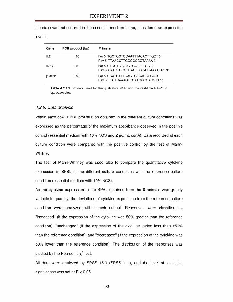
EXPERIMENT 2
92
the six cows and cultured in the essential medium alone, considered as expression
level 1.
Gene PCR product (bp) Primers
IL2 100 For 5’ TGCTGCTGGAATTTACAGTTGCT 3’ Rev 5’ TTAACCTTGGGCGCGTAAAA 3’ INFγ 103 For 5’ CTGCTCTGTGGGCTTTTGG 3’ Rev 5’ CATCTGGGCTACTTGCATTAAAATAC 3’
β-actin 183 For 5’ CCATCTATGAGGGTCACGCGC 3’ Rev 5’ TTCTCAAAGTCCAAGGCCACGTA 3’
Table 4.2.4.1. Primers used for the qualitative PCR and the real-time RT-PCR; bp: basepairs.
4.2.5. Data analysis
Within each cow, BPBL proliferation obtained in the different culture conditions was
expressed as the percentage of the maximum absorbance observed in the positive
control (essential medium with 10% NCS and 2 µg/mL conA). Data recorded at each
culture condition were compared with the positive control by the test of Mann-
Whitney.
The test of Mann-Whitney was used also to compare the quantitative cytokine
expression in BPBL in the different culture conditions with the reference culture
condition (essential medium with 10% NCS).
As the cytokine expression in the BPBL obtained from the 6 animals was greatly
variable in quantity, the deviations of cytokine expression from the reference culture
condition were analyzed within each animal. Responses were classified as
''increased'' (if the expression of the cytokine was 50% greater than the reference
condition), ''unchanged'' (if the expression of the cytokine varied less than ±50%
than the reference condition), and ''decreased'' (if the expression of the cytokine was
50% lower than the reference condition). The distribution of the responses was
studied by the Pearson’s χ2-test.
All data were analyzed by SPSS 15.0 (SPSS Inc.), and the level of statistical
significance was set at P < 0.05.
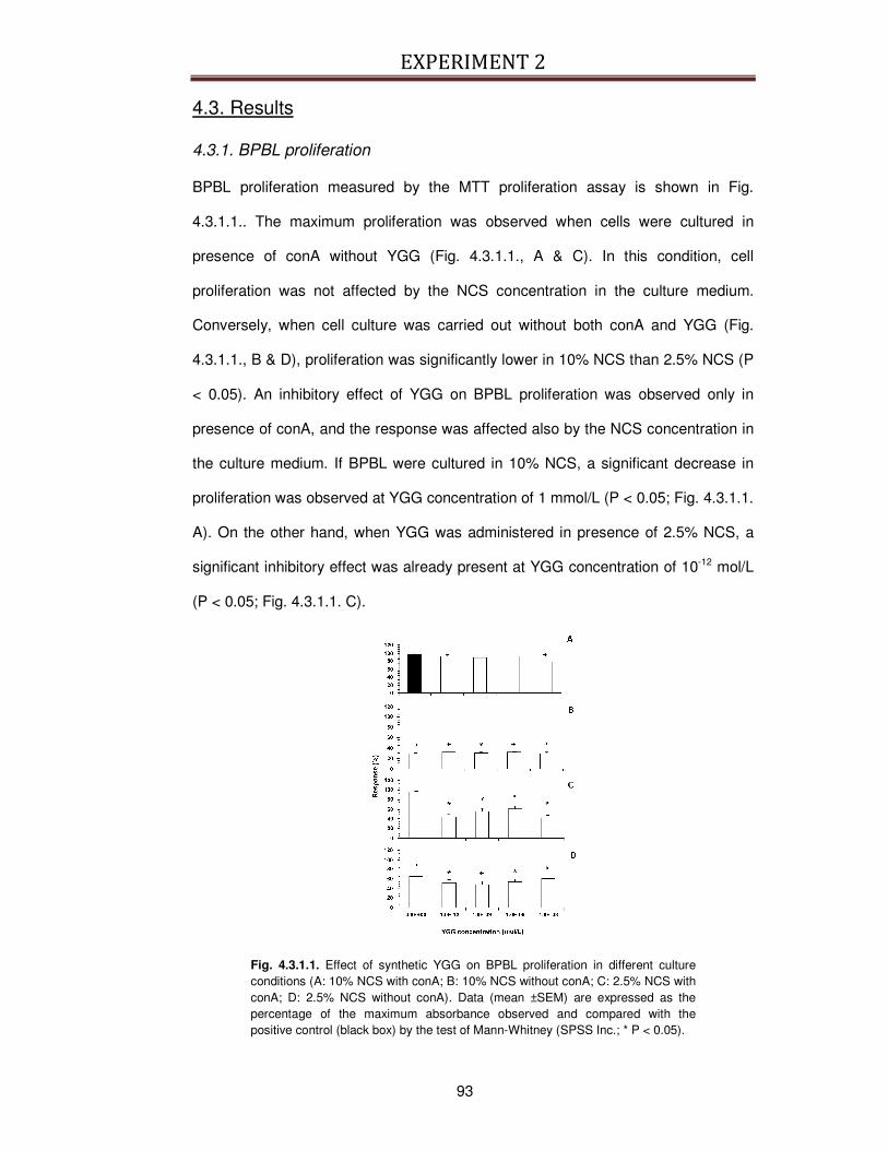
EXPERIMENT 2
93
4.3. Results
4.3.1. BPBL proliferation
BPBL proliferation measured by the MTT proliferation assay is shown in Fig.
4.3.1.1.. The maximum proliferation was observed when cells were cultured in
presence of conA without YGG (Fig. 4.3.1.1., A & C). In this condition, cell
proliferation was not affected by the NCS concentration in the culture medium.
Conversely, when cell culture was carried out without both conA and YGG (Fig.
4.3.1.1., B & D), proliferation was significantly lower in 10% NCS than 2.5% NCS (P
< 0.05). An inhibitory effect of YGG on BPBL proliferation was observed only in
presence of conA, and the response was affected also by the NCS concentration in
the culture medium. If BPBL were cultured in 10% NCS, a significant decrease in
proliferation was observed at YGG concentration of 1 mmol/L (P < 0.05; Fig. 4.3.1.1.
A). On the other hand, when YGG was administered in presence of 2.5% NCS, a
significant inhibitory effect was already present at YGG concentration of 10-12 mol/L
(P < 0.05; Fig. 4.3.1.1. C).
Fig. 4.3.1.1. Effect of synthetic YGG on BPBL proliferation in different culture conditions (A: 10% NCS with conA; B: 10% NCS without conA; C: 2.5% NCS with conA; D: 2.5% NCS without conA). Data (mean ±SEM) are expressed as the percentage of the maximum absorbance observed and compared with the positive control (black box) by the test of Mann-Whitney (SPSS Inc.; * P < 0.05).
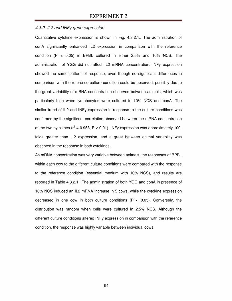
EXPERIMENT 2
94
4.3.2. IL2 and INFγ gene expression
Quantitative cytokine expression is shown in Fig. 4.3.2.1.. The administration of
conA significantly enhanced IL2 expression in comparison with the reference
condition (P < 0.05) in BPBL cultured in either 2.5% and 10% NCS. The
administration of YGG did not affect IL2 mRNA concentration. INFγ expression
showed the same pattern of response, even though no significant differences in
comparison with the reference culture condition could be observed, possibly due to
the great variability of mRNA concentration observed between animals, which was
particularly high when lymphocytes were cultured in 10% NCS and conA. The
similar trend of IL2 and INFγ expression in response to the culture conditions was
confirmed by the significant correlation observed between the mRNA concentration
of the two cytokines (r2 = 0.953, P < 0.01). INFγ expression was approximately 100-
folds greater than IL2 expression, and a great between animal variability was
observed in the response in both cytokines.
As mRNA concentration was very variable between animals, the responses of BPBL
within each cow to the different culture conditions were compared with the response
to the reference condition (essential medium with 10% NCS), and results are
reported in Table 4.3.2.1.. The administration of both YGG and conA in presence of
10% NCS induced an IL2 mRNA increase in 5 cows, while the cytokine expression
decreased in one cow in both culture conditions (P < 0.05). Conversely, the
distribution was random when cells were cultured in 2.5% NCS. Although the
different culture conditions altered INFγ expression in comparison with the reference
condition, the response was highly variable between individual cows.
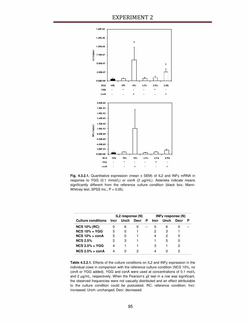
EXPERIMENT 2
95
Fig. 4.3.2.1. Quantitative expression (mean ± SEM) of IL2 and INFγ mRNA in response to YGG (0.1 mmol/L) or conA (2 µg/mL). Asterisks indicate means significantly different from the reference culture condition (black box; Mann-Whitney test; SPSS Inc.; P < 0.05).
IL2 response (N) INFγ response (N) Culture conditions Incr Unch Decr P Incr Unch Decr P
NCS 10% (RC) 0 6 0 -- 0 6 0 -- NCS 10% + YGG 5 0 1 2 3 1 NCS 10% + conA 5 0 1 4 2 0 NCS 2.5% 2 3 1 1 5 0
NCS 2.5% + YGG 4 1 1 3 1 2
NCS 2.5% + conA 4 0 2 4 0 2
Table 4.3.2.1. Effects of the culture conditions on IL2 and INFγ expression in the individual cows in comparison with the reference culture condition (NCS 10%, no conA or YGG added). YGG and conA were used at concentrations of 0.1 mol/L and 2 µg/mL, respectively. When the Pearson’s χ2 test in a row was significant, the observed frequencies were not casually distributed and an effect attributable to the culture condition could be postulated. RC: reference condition; Incr: increased; Unch: unchanged; Decr: decreased.
EXPERIMENT 2
96
4.4. Discussion
In this work, the possibility to use the bovine as an alternative source of lymphocytes
has been explored to develop a bioassay to test immunoactive peptides, as blood
samples from animals slaughtered for commercial meat production are available in
practically unlimited amounts. This would allow avoiding the use of laboratory
animals and overcoming the need to recruit human volunteers.
As YGG and YG are immunoactive peptides resulting from both enkephalin [154,
185] and milk protein [134] cleavage, they may represent an interesting model for
functional immunoactive peptides of food origin, providing that they can be released
during digestion and cross the intestinal barrier. The release of YGG from
enkephalins can be achieved by the action of enzymes such as aminopeptidase N
(EC 3.4.11.2), peptidyl-dipeptidase A (EC 3.4.15.1) and endopeptidase 24.11 (EC
3.4.24.11), which can be secreted by or are associated to immune cells and other
tissues [316, 317]. Interestingly, those enzymes are expressed in the brush border
surface of the human and rat enterocytes [191, 318], suggesting that YGG
encrypted in milk proteins could be released. Furthermore, several studies revealed
optimistic perspectives about the intestinal absorption of small peptides, in particular
di- and tri-peptides, as there are indications that they can escape from the action of
brush-border and cytoplasmic peptidases [186, 201, 244, 319-321], and their
transepithelial transport may be achieved by carrier-mediated transport through
PepT1 [322] or by paracellular route [186, 321].
As YG seems to be the most active form [134], while YGG is thought to be the main
product of enkephalin degradation and less susceptible to protease attack [323,
324], the latter has been used as a model of potentially absorbable immunoactive
peptide. If the peptide reached the circulation it could interact with the cells of the
immune system and exert its function(s), although plasma/serum components could
affect YGG activity.

EXPERIMENT 2
97
To highlight the effect of serum on YGG activity, BPBL has been cultured in
presence of two concentrations of NCS, and it has been observed that higher NCS
concentrations showed an inhibitory effect on cell proliferation per se, which was
always abolished by the addition of conA in the culture medium. Conversely, no
effect of serum concentration on quantitative cytokine expression was detectable,
even though other works indicated that variations in NCS concentration could affect
cytokine expression in lymphocytic leukemia cells [325] and in a macrophagic cell
line, where it could depress TNFα production [326].
Although the effects of YGG on the cells of the immune system has been proven
also in vivo [154, 185], its mechanism of action is far from being fully elucidated. At
present, only few papers [134, 155, 157] examined the effects of YGG in vitro on
lymphocytes using different experimental approaches. Sizemore and colleagues
[155] studied the effects of YG and YGG on conA-induced regulatory T cell activity
on cell proliferation. They found that both peptides increased in vitro proliferation of
T cells stimulated by conA, and YG showed the greatest biological activity.
Moreover, those authors observed a biphasic effect as YGG stimulated proliferation
at lower concentrations (10-13 – 10-14 mol/L) and inhibited proliferation at higher
concentrations.
More recently, Kayser and Meisel [134] stimulated with YGG human peripheral
blood lymphocytes previously activated with conA and estimated cell proliferation by
BrdU incorporation. Proliferation was only slightly stimulated by YGG in comparison
with the dipeptide YG, and the maximum increase in lymphocyte proliferation
induced by YGG was about 20-30% of the control (culture medium only). In addition,
the stimulatory effect of YGG on cell proliferation was abolished at higher
concentrations (10-4 - 10-5 mol/L) of peptide added. Unfortunately, that paper did not
give information about serum effects on YGG activity.
In the present work, YGG did show neither mitogenic activity, as it did not alter cell
proliferation if added alone to the cell culture, nor additive action to the pro-
EXPERIMENT 2
98
proliferative effect of conA over the concentration interval under examination. Likely,
no pro-proliferative effect of YGG could be detected because peptide concentrations
lower than 10-12 mol/L were not tested, which likely are pro-proliferative [327].
However, an inhibitory effect of YGG on conA-induced BPBL proliferation was
observed, as the peptide decreased proliferation by 20-60%, depending on the
concentration used. In addition, NCS concentrations clearly modulated the BPBL
proliferative response to YGG. In fact, lymphocytes were more sensitive to YGG at
lower NCS concentration, indicating an inhibitory action of serum on the peptide
activity. This is in agreement with previous observations that enkephalins and YGG
can be rapidly hydrolyzed/inactivated in human plasma [328-331], and plasma
proteins can bind enkephalins and their related peptides [327, 332] rendering them
unable to exert their effects.
The majority of T cells responds to and produces IL2 upon activation. Piva et al.
[157] reported that both YG and YGG could affect the expression of IL2, IL4 and
INFγ in murine splenocytes activated with suboptimal concentration of conA in a
serum-free culture system. Those authors observed that both peptides stimulated
INFγ protein production at very low concentrations (10-13 mol/L) and inhibited both
INFγ and IL2 at higher concentrations (10-7 - 10-3 mol/L), while no enhancing effect
on IL2 secretion could be detected.
In the present study, the ability of YGG to affect IL2 and INFγ expression was
studied using 10-3 mol/L YGG, because an effect on BPBL proliferation could always
be observed at that peptide concentration. To summarize, when BPBL were
activated with conA, they responded increasing both proliferation and cytokine
expression, IL2 in particular. On the contrary, no significant YGG effect on IL2 and
INFγ mRNA concentrations could be seen, although a slight IL2 mRNA increase in 5
out 6 animals was detected if the peptide was administered in presence of 10%
NCS. It is possible that the inhibitory YGG action on cytokine expression was

EXPERIMENT 2
99
suppressed/masked by the presence of NCS in the culture medium, and it might be
observed only in a serum-free culture system.
It was hypothesized that YGG and YG could exert their suppressive effects through
a yet unidentified binding site that selectively binds those peptides with low affinity
[157].
In conclusion, the anti-proliferative effect of YGG could be observed in vitro also in
BPBL, despite the presence of serum in the culture medium. However, serum
concentration significantly influenced the assay outcome, as proliferation of conA-
activated BPBL was inhibited in a manner inversely related to NCS concentration.
On the other hand, high YGG concentrations did not inhibit the synthesis of IL2 and
INFγ mRNA. The use of bovine lymphocyte culture as a bioassay to evaluate the
action of immunomodulatory substances needs to be further validated examining
more culture conditions and, perhaps, selecting different lymphocyte populations. In
this respect, it is important to consider that peripheral lymphocytes may not be the
target for immunoactive peptides introduced by the diet. In fact, YGG present in the
gut can have a good chance to exert its biological activity without reaching the
circulation, as it may be hypothesized that the peptide can be transferred to the
gastrointestinal-associated lymphoid tissue (GALT) at the level of Payer’s patches
by Antigen-Presenting Cells, where it may exert its putative effects at the serosa
level by influencing cytokine release [333].
4.5. Take-home message
The present work demonstrated that the bioactive peptide YGG had an
immunomodulatory activity and, in particular, it modulated the proliferation of
mitogen-activated bovine lymphocytes. Kayser and Meisel [134] previously
demonstrated that YGG peptide modulated the proliferation of lymphocytes of other
species, more specifically human lymphocytes. In both cases, the effects of YGG
EXPERIMENT 2
100
were evaluated on isolated lymphocytes maintained in culture. It is possible that in
the serum added to the culture medium some unidentified factors affect the activity
of YGG. In the present work, to acquire better knowledge of the role of NCS, the
effect of YGG on lymphocytes proliferation has been evaluated at two specific
serum concentrations in the culture medium. The next step would be to characterize
YGG effects when serum is not present in the culture medium.
Another important information that can be acquired from the present study is about
the stability of YGG to serum peptidases obtained incubating the peptide with
medium added with serum. In fact, serum peptidases may influence the effects of
YGG during proliferation test but also may contribute to determine the possibility of
the peptide to reach intact its target site, once absorbed and circulating into the
body.
The present work explored also the possibility to use the bovine lymphocytes to test
immunomodulatory peptides. The preliminary results obtained are encouraging but a
deeper characterization of the isolated lymphocytes. In particular, it would be useful
to characterize the cytokines that they express when they are activated, and the
response to the different concentrations of serum in the culture medium, because it
would help to understand the molecular mechanisms of these bioactive peptides.

EXPERIMENT 3
101
EXPERIMENT 3: Study of the bioactive properties and the transport of the peptide β-CN (193-209), a 17-residues peptide of bovine β-casein, through Caco-2 monolayers 1
5.1. Introduction
Milk proteins are a source of peptides that exhibit numerous bioactivities including
antihypertensive, opiate, immunomodulatory, antimicrobial, antioxidant or mineral-
binding activities [15, 48, 77, 334, 335]. Among those, the β-CN (193–209) peptide
is released from the C-terminal end of β-casein by hydrolysis with pepsin. This
peptide was isolated and identified from yoghurt and fermented milks as well as
several types of cheese [60, 187, 188]. It is a 17 residues long peptide with the
amino acid sequence Tyr-Gln-Glu-Pro-Val-Leu-Gly-Pro-Val-Arg-Gly-Pro-Phe-Pro-
Ile-Ile-Val. This peptide displays immunomodulatory properties and shows mitogenic
activity on primed lymph node cells and unprimed rat spleen cells [147]. It manifests
chemotactic activity on L14 lymphoblastoid cell line [189], and enhances
phagocytosis in rat macrophages [148, 190].
To exert their biological activity, some peptides have to cross the gastrointestinal
barrier, and reach the circulation and target sites in an active form [261]. Resistance
to enzymatic degradation and transport through intestinal cells are the two important
factors influencing the bioavailability of orally ingested peptides. There are some
distinctive features determining the possibility of a peptide to be absorbed intact
through the intestinal epithelium, such as its molecular mass, hydrophobicity, charge
or tendency to aggregate [336, 337]. Interestingly, the presence of 4 proline
residues within the sequence can protect the long β-CN (193–209) peptide from the
action of peptidases. As a consequence, this peptide appears as a good candidate
for crossing the intestinal barrier in an intact bioactive form. The main routes 1 Part of this experiment has been accepted for publication by Molecular Nutrition & Food Research with the
following title “The (109-203) 17-residues of β-casein is transported through Caco-2 monolayer”, written by Regazzo D., Mollé D., Gabai G., Tomé D., Dupont D., Léonil J. and Boutrou R.
EXPERIMENT 3
102
recognized for transepithelial absorption of peptide in the gut include the PepT1
transporter-mediated transport for di- and tri-peptides, the paracellular passive
transport via tight junctions, the transcellular passive diffusion and transcytosis that
is a transcellular route involving endocytotic uptake, intracellular transport via
transcytotic vesicles and basolateral secretion [338].
The aim of this study was to determine the sensitivity of the β-CN (193–209) peptide
to hydrolysis by brush border enzymes and its transepithelial transport across Caco-
2 cell monolayer as a model of intestinal epithelium. The pathway of transepithelial
transport was investigated by using selective inhibitors of the different routes,
including the dipeptide Gly-Pro that competitively inhibits the peptide transporter
PepT1 [336], cytochalasin D that opens tight junctions by altering the cytoskeletal
structure [339] and increasing the passive paracellular route, and wortmannin as an
inhibitor of transcytosis [340]. In addition the effects of this immunomodulatory
peptide on the viability and tight junction stability of Caco-2 cells was investigated to
better characterize its biological activities.
5.2. Materials and Methods
5.2.1. Chemicals and Reagents
Dulbecco's modified Eagle's medium (DMEM), non-essential amino acids (NEAA),
gentamycin sulphate, Hank's balanced salt solution (HBSS) and phosphate buffered
saline (PBS) were purchased from Lonza, Switzerland. Gibco-Invitrogen (United
Kindom) supplied L-Glutamine (L-Glu) and trypsine-EDTA. Fetal calf serum (FCS)
was purchased from Dutscher, France. Sigma–Aldrich, (France) supplied
cytochalasin D, dimetilsulfoxide (DMSO), Glycil-Proline dipeptide (Gly-Pro), glycine,
glucose, HCl, hydroxyethyl piperazine ethane sulphonic acid (HEPES), mannitol,
neutral red powder, para-nitrophenyl phosphate, trinitrobenzenesulfonic acid (TNBS)
and Triton X100. Wortmannin was obtained from LC Laboratories, Massachusetts,
EXPERIMENT 3
103
USA. Acetonitrile, acetic acid ethanol, 2-Amino-(hydroxymethyl)-1,3-propanediol
(TRIS) and trifluoroacetic acid (TFA) were purchased from Fluka, France.
5.2.2. Preparation of β-CN (193-209)
The peptide was obtained in a purified form as previously described [341].
Conditions for hydrolyzing β-CN were slightly modified for β-CN concentration (5
g/L), molar ratio chymosin/β-CN (1/8000) and duration of hydrolysis (150 min). Then,
the reaction was stopped by heat inactivation of the enzyme (80°C, 15 min). The pH
of the mixture was subsequently adjusted to 4.6 with 1 mol/L HCl to precipitate and
remove by centrifugation (7000 x g for 20 min) whole casein and its large fragments.
After readjusting the pH to 6.5, the supernatant was ultra filtered (Spiral-wound UF
cartridge S10T3 MWCO 3 KDa; Amicon, Lexington, Massachusetts, USA) and the
ultra filtrate was concentrated with a membrane (Filtron membrane 1 KDa) and then
freeze-dried. The peptide, identified by electrospray mass spectrometry (ESI/MS),
was obtained with a purity of 98% estimated by RP-HPLC-ESI/MS as described in
Paragraph 5.2.7.
5.2.3. Cell Culture
Caco-2 cells were obtained from the American Type Culture Collection (Rockville,
Maryland, USA). Cells were cultured in DMEM supplemented with 20% FCS, 1%
NEAA, 2 mmol/L L-Glu and 25 µg/mL gentamycin sulphate. They were incubated at
37°C in humidified atmosphere containing 5% CO2. The monolayer became
confluent 4-5 days after seeding 3·106 cells/flasks (75 cm2 flasks, Greiner Bio-one,
France), and the cells were subcultured at split ratio of 1:5 by trypsinization (0.5%
trypsin and 0.05% EDTA). The medium was changed every second day. The cells
used in this study were at passages 35-45. For transport studies, cells were seeded
in cell culture inserts with Anopore membranes (0.2 µm pore sizes; 25 mm
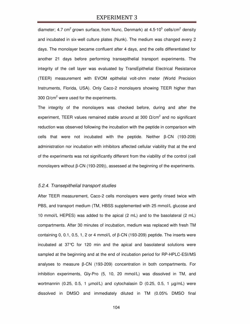
EXPERIMENT 3
104
diameter; 4.7 cm2 grown surface, from Nunc, Denmark) at 4.5·105 cells/cm2 density
and incubated in six-well culture plates (Nunk). The medium was changed every 2
days. The monolayer became confluent after 4 days, and the cells differentiated for
another 21 days before performing transepithelial transport experiments. The
integrity of the cell layer was evaluated by TransEpithelial Electrical Resistance
(TEER) measurement with EVOM epithelial volt-ohm meter (World Precision
Instruments, Florida, USA). Only Caco-2 monolayers showing TEER higher than
300 Ω/cm2 were used for the experiments.
The integrity of the monolayers was checked before, during and after the
experiment, TEER values remained stable around at 300 Ω/cm2 and no significant
reduction was observed following the incubation with the peptide in comparison with
cells that were not incubated with the peptide. Neither β-CN (193-209)
administration nor incubation with inhibitors affected cellular viability that at the end
of the experiments was not significantly different from the viability of the control (cell
monolayers without β-CN (193-209)), assessed at the beginning of the experiments.
5.2.4. Transepithelial transport studies
After TEER measurement, Caco-2 cells monolayers were gently rinsed twice with
PBS, and transport medium (TM, HBSS supplemented with 25 mmol/L glucose and
10 mmol/L HEPES) was added to the apical (2 mL) and to the basolateral (2 mL)
compartments. After 30 minutes of incubation, medium was replaced with fresh TM
containing 0, 0.1, 0.5, 1, 2 or 4 mmol/L of β-CN (193-209) peptide. The inserts were
incubated at 37°C for 120 min and the apical and basolateral solutions were
sampled at the beginning and at the end of incubation period for RP-HPLC-ESI/MS
analyses to measure β-CN (193-209) concentration in both compartments. For
inhibition experiments, Gly-Pro (5, 10, 20 mmol/L) was dissolved in TM, and
wortmannin (0.25, 0.5, 1 µmol/L) and cytochalasin D (0.25, 0.5, 1 µg/mL) were
dissolved in DMSO and immediately diluted in TM (0.05% DMSO final

EXPERIMENT 3
105
concentration). The cell monolayers were incubated 30 min with the inhibitors or
with 0.05% DMSO, as a control, before the peptide transport experiments. During
and at the end of the experiments, TEER was measured and data were recorded
only from experiments in which TEER was higher than 250 Ω/cm2. To exclude that
addition of β-CN (193-209) and/or inhibitors could be toxic for the cells, cellular
viability was assessed at the end of each experiment using the vital dye neutral red,
as described in the Paragraph 5.2.5.
5.2.5. Effects of β-CN (193-209) on cellular viability
To assess if peptide addition could be toxic for the cells, cellular viability was
evaluated at the end of each experiment using the vital dye neutral red (NR). NR is
a weak cationic dye that diffuses readily across plasma and organelle membranes,
accumulating in the lysosomes. The principle of the assay is based on the fact that
the loss of membrane integrity induced by test agents results in decreased retention
of NR (quantification of NR at 540 nm). Damaged or dead cells thus appear
unstained in comparison with healthy control cells. Similarly, lower absorbance after
NR extraction is an indication of reduced cellular viability [342]. The applied protocol
is based on the NR uptake assay first described by Borenfreund and Puerner [343].
Briefly, TM used for the incubation was removed, the cells were washed twice with
sterile PBS and fresh TM was replaced. Then Neutral Red Solution (0.33% in PBS,
w/v) was added to the medium in an amount equal to 10% of the medium volume
and cells were allowed to incubate for 120 min at 37 °C. At the end of the incubation
period, the medium was carefully removed, the cells quickly rinsed twice with sterile
PBS. The incorporated dye was then solubilized in a volume of Solubilization
Solution (1% acetic acid, 50% ethanol, 49% milliQ water) equal to the original
volume of culture medium. The cultures were allowed to stand for 10 minutes at
room temperature, enhancing the dye solubilization by gentle stirring in a rotatory
shaker. The absorbance was measured at a wavelength of 540 nm with background
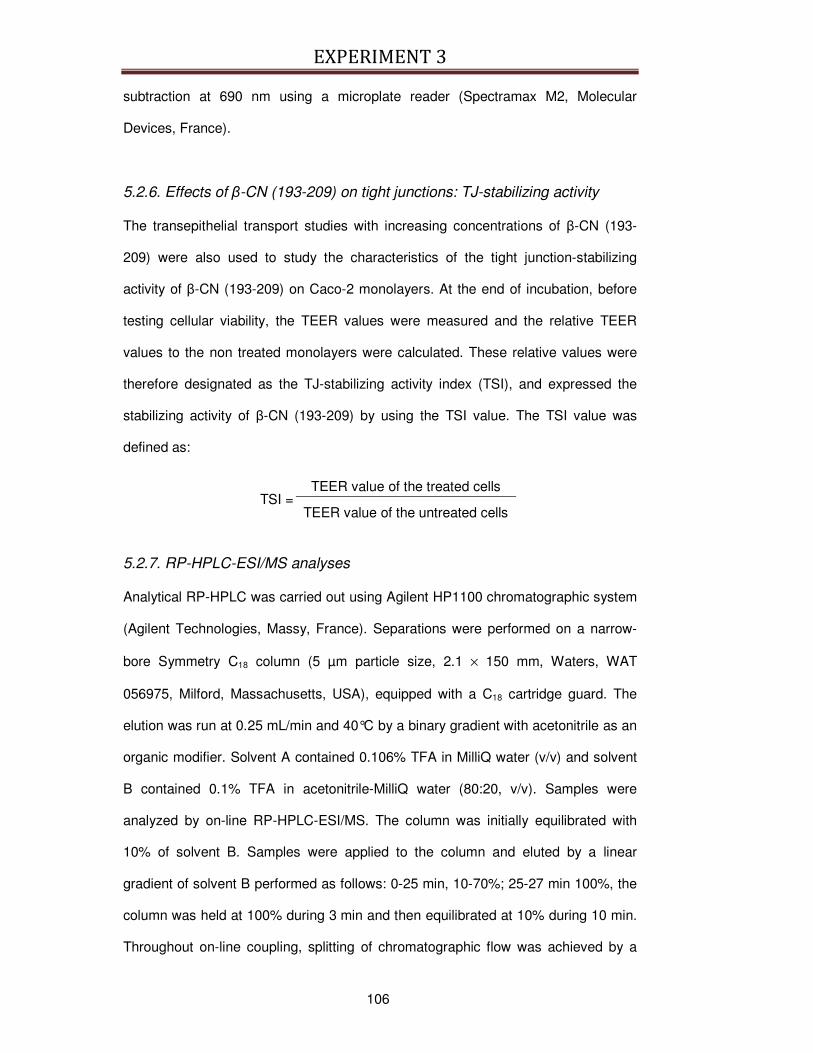
EXPERIMENT 3
106
subtraction at 690 nm using a microplate reader (Spectramax M2, Molecular
Devices, France).
5.2.6. Effects of β-CN (193-209) on tight junctions: TJ-stabilizing activity
The transepithelial transport studies with increasing concentrations of β-CN (193-
209) were also used to study the characteristics of the tight junction-stabilizing
activity of β-CN (193-209) on Caco-2 monolayers. At the end of incubation, before
testing cellular viability, the TEER values were measured and the relative TEER
values to the non treated monolayers were calculated. These relative values were
therefore designated as the TJ-stabilizing activity index (TSI), and expressed the
stabilizing activity of β-CN (193-209) by using the TSI value. The TSI value was
defined as:
TSI = TEER value of the treated cells
TEER value of the untreated cells
5.2.7. RP-HPLC-ESI/MS analyses
Analytical RP-HPLC was carried out using Agilent HP1100 chromatographic system
(Agilent Technologies, Massy, France). Separations were performed on a narrow-
bore Symmetry C18 column (5 µm particle size, 2.1 × 150 mm, Waters, WAT
056975, Milford, Massachusetts, USA), equipped with a C18 cartridge guard. The
elution was run at 0.25 mL/min and 40°C by a binary gradient with acetonitrile as an
organic modifier. Solvent A contained 0.106% TFA in MilliQ water (v/v) and solvent
B contained 0.1% TFA in acetonitrile-MilliQ water (80:20, v/v). Samples were
analyzed by on-line RP-HPLC-ESI/MS. The column was initially equilibrated with
10% of solvent B. Samples were applied to the column and eluted by a linear
gradient of solvent B performed as follows: 0-25 min, 10-70%; 25-27 min 100%, the
column was held at 100% during 3 min and then equilibrated at 10% during 10 min.
Throughout on-line coupling, splitting of chromatographic flow was achieved by a

EXPERIMENT 3
107
low dead volume tee with 85% of the flow directed to the UV detector and 15% to
the mass spectrometer. This split allows a perfect superposition between UV and
TIC (Total Ionization Current) detection. Peaks were detected both by UV
absorbance at 214 nm and peptide mass spectrometry by TIC.
The β-CN (193–209) peptide was quantified in accordance to a standard curve,
established with chosen quantities (from 0.053 nmol to 1.063 nmol) of purified
peptide β-CN (193–209). β-CN (193–209) quantity (x) in apical and basolateral
solutions was calculated with the equation y=bx+c, where y is the UV absorbance at
214 nm, c is the y-axis intercept and b the slope of a standard curve.
The proteolysis of the peptide was analyzed by LC-MS. The mass spectrometer
(API III+ SCIEX, Thornhill, Ontario, Canada) comprises a triple quadrupole equipped
with an atmospheric pressure ionization source. Analysis was carried out in positive
detection mode. A 75 µm sprayer was usually set at 4800 V for generated multiply-
charged ions and orifice set between 60 to 90 V depending on experiments. The
nebulizer pressure was set around 0.5 MPa and the curtain gas set to 1.2 L/min.
The instrument mass-to-charge (m/z) scale was calibrated with polypropylene
glycols. All peptide mass spectra were obtained from the average signal of multiple
scans. Each scan was acquired over the range of m/z values from 500 to 2000
using a step size of 0.5 Da and a dwell time of 0.5 ms. The measured masses were
matched with predicted enzymatic fragments by using the software BioMultiview
1.3.1 (MDS Perkin Elmer Sciex, Thornhill, Canada).
5.2.8. Assessment of β-CN (193-209) hydrolysis
Hydrolysis of β-CN (193-209) was determined by measuring free amino groups (-
NH2 groups) with trinitrobenzenesulfonic acid (TNBS) as described by Boutrou et al.
[344], following 1:4 dilution in distilled water. Briefly, the supernatant (10 µL) was
added to 100 µL of potassium borate (1 mol/L, pH 9.2) and 40 µL of TNBS (1.2 g/L).
After incubation (1 h, 37°C), the absorbance was measured at 405 nm using a
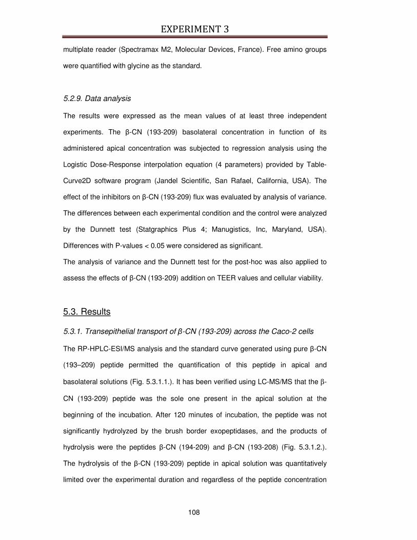
EXPERIMENT 3
108
multiplate reader (Spectramax M2, Molecular Devices, France). Free amino groups
were quantified with glycine as the standard.
5.2.9. Data analysis
The results were expressed as the mean values of at least three independent
experiments. The β-CN (193-209) basolateral concentration in function of its
administered apical concentration was subjected to regression analysis using the
Logistic Dose-Response interpolation equation (4 parameters) provided by Table-
Curve2D software program (Jandel Scientific, San Rafael, California, USA). The
effect of the inhibitors on β-CN (193-209) flux was evaluated by analysis of variance.
The differences between each experimental condition and the control were analyzed
by the Dunnett test (Statgraphics Plus 4; Manugistics, Inc, Maryland, USA).
Differences with P-values < 0.05 were considered as significant.
The analysis of variance and the Dunnett test for the post-hoc was also applied to
assess the effects of β-CN (193-209) addition on TEER values and cellular viability.
5.3. Results
5.3.1. Transepithelial transport of β-CN (193-209) across the Caco-2 cells
The RP-HPLC-ESI/MS analysis and the standard curve generated using pure β-CN
(193–209) peptide permitted the quantification of this peptide in apical and
basolateral solutions (Fig. 5.3.1.1.). It has been verified using LC-MS/MS that the β-
CN (193-209) peptide was the sole one present in the apical solution at the
beginning of the incubation. After 120 minutes of incubation, the peptide was not
significantly hydrolyzed by the brush border exopeptidases, and the products of
hydrolysis were the peptides β-CN (194-209) and β-CN (193-208) (Fig. 5.3.1.2.).
The hydrolysis of the β-CN (193-209) peptide in apical solution was quantitatively
limited over the experimental duration and regardless of the peptide concentration

EXPERIMENT 3
109
(Fig. 5.3.1.3A. and Fig. 5.3.1.3B.). When the peptide was added at 2 mmol/L, a 10%
decrease in its concentration in the apical compartment was observed after 2 h
incubation, and the peptide left remained intact. For higher concentrations, the
hydrolysis was less than 10%. RP-HPLC-ESI/MS analysis of basolateral solution
showed that the β-CN (193-209) peptide and its two derived fragments were
absorbed intact through Caco-2 monolayer. After 120 min incubation in the apical
compartment at the milli molar range, the β-CN (193-209) peptide appeared in
basolateral compartment at the micro molar range with concentration values
following a saturable pattern (Fig. 5.3.1.4.), described by a sigmoidal curve.
Fig. 5.3.1.1. Identification and quantification of β-CN (193-209) by RP-HPLC-ESI/MS analysis. A. Identification and estimation of β-CN (193-209) purity level are shown on the spectrum and on the TIC graph. β-CN (193-209) was added in the apical compartment at 2 mmol/L and apical solution immediately analyzed. B. Quantification of β-CN (193-209) in apical and basolateral solutions using a five-point calibration curve of pure β-CN (193-209) as standard analyzed by RP-HPLC-ESI/MS (see Paragraph 5.2.7).
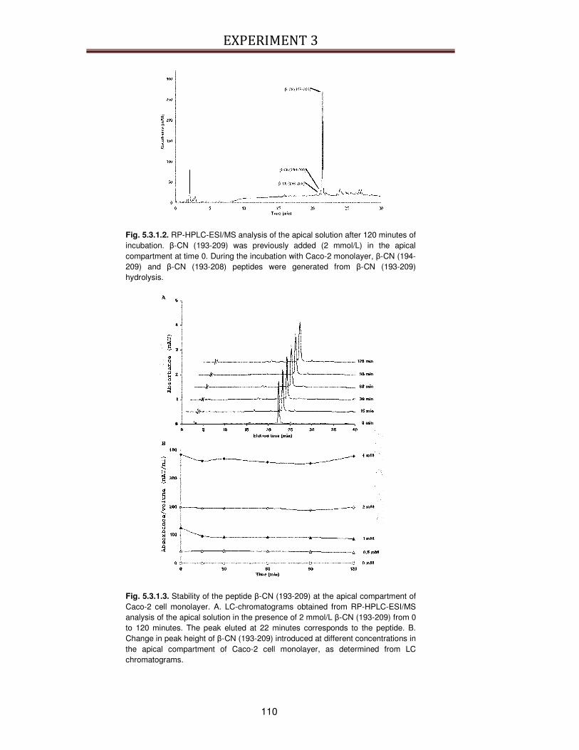
EXPERIMENT 3
110
Fig. 5.3.1.2. RP-HPLC-ESI/MS analysis of the apical solution after 120 minutes of incubation. β-CN (193-209) was previously added (2 mmol/L) in the apical compartment at time 0. During the incubation with Caco-2 monolayer, β-CN (194-209) and β-CN (193-208) peptides were generated from β-CN (193-209) hydrolysis.
Fig. 5.3.1.3. Stability of the peptide β-CN (193-209) at the apical compartment of Caco-2 cell monolayer. A. LC-chromatograms obtained from RP-HPLC-ESI/MS analysis of the apical solution in the presence of 2 mmol/L β-CN (193-209) from 0 to 120 minutes. The peak eluted at 22 minutes corresponds to the peptide. B. Change in peak height of β-CN (193-209) introduced at different concentrations in the apical compartment of Caco-2 cell monolayer, as determined from LC chromatograms.

EXPERIMENT 3
111
Fig. 5.3.1.4. Concentration dependence transport of β-CN (193-209) across Caco-2 monolayer. Different amounts of peptide were incubated in apical compartment and β-CN (193-209) was evaluated in basolateral solution after 120 minutes incubation at 37°C. Quantification was obtained as described in Paragraph 5.2.7.
5.3.2. Influence of Gly-Pro, Cytochalasin D and wortmannin on β-CN (193-
209) transport
To evaluate the pathway of the transepithelial transport of β-CN (193-209), the
effect of some inhibitors on the apical to basolateral flux of β-CN (193-209) was
tested (Fig. 5.3.2.1.). The transport of β-CN (193-209) was not significantly
decreased by Gly-Pro (applied from 5 to 20 mmol/L) that competitively inhibits the
peptide transporter PepT1. In the range from 0.25 to 1 µg/mL, the treatment with
cytochalasin D, a tight junctions disruptor, reduced TEER values approximately of
20%, indicating that paracellular route was similarly expanded regardless the
concentration of the β-CN (193-209). Nevertheless, the presence of cytochalasin D
at 0.25, 0.5 and 1 µg/mL did not significantly alter apical to basolateral flux at any
concentration used. On the contrary, the addition of the inhibitor of transcytosis
wortmannin in the range from 0.25 to 1 µmol/L significantly (P < 0.05) reduced the
flux of β-CN (193-209) through the Caco-2 monolayer and an average 53%
decrease of transport was determined (Fig. 5.3.2.1.).
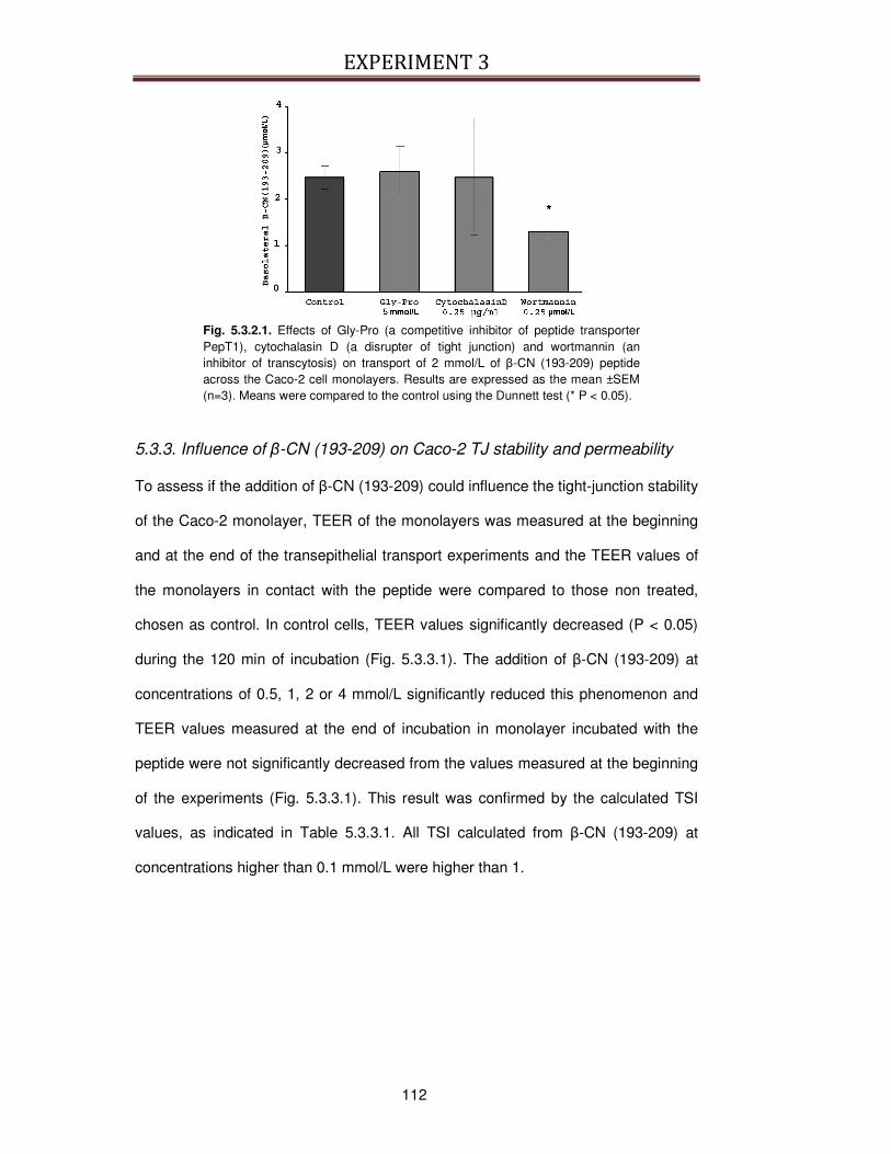
EXPERIMENT 3
112
Fig. 5.3.2.1. Effects of Gly-Pro (a competitive inhibitor of peptide transporter PepT1), cytochalasin D (a disrupter of tight junction) and wortmannin (an inhibitor of transcytosis) on transport of 2 mmol/L of β-CN (193-209) peptide across the Caco-2 cell monolayers. Results are expressed as the mean ±SEM (n=3). Means were compared to the control using the Dunnett test (* P < 0.05).
5.3.3. Influence of β-CN (193-209) on Caco-2 TJ stability and permeability
To assess if the addition of β-CN (193-209) could influence the tight-junction stability
of the Caco-2 monolayer, TEER of the monolayers was measured at the beginning
and at the end of the transepithelial transport experiments and the TEER values of
the monolayers in contact with the peptide were compared to those non treated,
chosen as control. In control cells, TEER values significantly decreased (P < 0.05)
during the 120 min of incubation (Fig. 5.3.3.1). The addition of β-CN (193-209) at
concentrations of 0.5, 1, 2 or 4 mmol/L significantly reduced this phenomenon and
TEER values measured at the end of incubation in monolayer incubated with the
peptide were not significantly decreased from the values measured at the beginning
of the experiments (Fig. 5.3.3.1). This result was confirmed by the calculated TSI
values, as indicated in Table 5.3.3.1. All TSI calculated from β-CN (193-209) at
concentrations higher than 0.1 mmol/L were higher than 1.
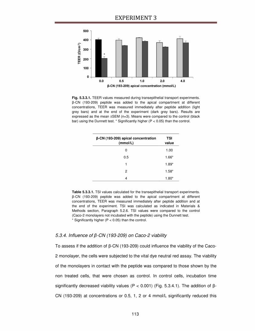
EXPERIMENT 3
113
Fig. 5.3.3.1. TEER values measured during transepithelial transport experiments. β-CN (193-209) peptide was added to the apical compartment at different concentrations, TEER was measured immediately after peptide addition (light grey bars) and at the end of the experiment (dark grey bars). Results are expressed as the mean ±SEM (n=3). Means were compared to the control (black bar) using the Dunnett test. * Significantly higher (P < 0.05) than the control.
β-CN (193-209) apical concentration (mmol/L)
TSI value
0 1.00
0.5 1.66*
1 1.89*
2 1.58*
4 1.80*
Table 5.3.3.1. TSI values calculated for the transepithelial transport experiments. β-CN (193-209) peptide was added to the apical compartment at different concentrations, TEER was measured immediately after peptide addition and at the end of the experiment. TSI was calculated as indicated in Materials & Methods section, Paragraph 5.2.6. TSI values were compared to the control (Caco-2 monolayers not incubated with the peptide) using the Dunnett test. * Significantly higher (P < 0.05) than the control.
5.3.4. Influence of β-CN (193-209) on Caco-2 viability
To assess if the addition of β-CN (193-209) could influence the viability of the Caco-
2 monolayer, the cells were subjected to the vital dye neutral red assay. The viability
of the monolayers in contact with the peptide was compared to those shown by the
non treated cells, that were chosen as control. In control cells, incubation time
significantly decreased viability values (P < 0.001) (Fig. 5.3.4.1). The addition of β-
CN (193-209) at concentrations or 0.5, 1, 2 or 4 mmol/L significantly reduced this

EXPERIMENT 3
114
phenomenon and viability values measured after 120 min of incubation in monolayer
incubated with the peptide were not significantly decreased from the viability values
measured at the beginning of the experiments (Fig. 5.3.4.1.).
Fig. 5.3.4.1. Viability values measured during transepithelial transport experiments. β-CN (193-209) peptide was added to the apical compartment at different concentrations, viability was measured immediately after peptide addition (light grey bars) and at the end of the experiment (dark grey bars). Results are expressed as the mean ±SEM (n=3). Means were compared to the control using the Dunnett test (* P < 0.001).
5.4. Discussion
The hypothesis that peptides escape digestion and are transported from the
intestinal lumen into blood circulation is gaining acceptance for small peptides,
mainly due to the growing number of studies describing the in vitro transepithelial
transport of bioactive peptides [186, 201, 202, 244, 261, 320, 321, 345-348]. In the
present study we demonstrate that the 17 residue β-CN (193-209) peptide and the
derived β-CN (193-208) and β-CN (194-209) peptides are transported across Caco-
2 cell monolayer, with the major contribution of the transcytosis mechanisms.
To exert its biological effects an ingested peptide must first resist intestinal
hydrolysis. To study the resistance of the β-CN (193-209) peptide to brush-border
membrane peptidases, Caco-2 cell monolayer has been used because, under
specific culture conditions, Caco-2 cells undergo a process of differentiation leading
to the expression of several morphological and functional characteristics of the

EXPERIMENT 3
115
enterocyte including the microvillus structure and of brush-border enzymes in the
apical membrane [227, 276]. About 10% of the β-CN (193-209) peptide were
hydrolyzed through the action of amino- and carboxy-peptidases present on the
apical membrane, but the β-CN (193-209) peptide and the two derived peptides, β-
CN (193-208) and β-CN (194-209), were not further hydrolyzed by brush-border
membrane exopeptidases. Moreover, the three peptides resisted the action of
intracellular peptidases. In general, due to their rapid hydrolysis by the brush border
or cytoplasmic peptidases, the bioavailability of 2 to 9 residues-peptides is extremely
low [186, 202, 244, 246, 261, 321, 346, 348]. The resistance of the β-CN (193-209)
peptide to the action of Caco-2 brush-border peptidases is possibly related to its
proline-rich sequence (4 proline residues on 17 residues), and other proline
containing peptides were found to be resistant to intestinal proteolysis [41, 257].
This finding is further confirmed by Savoie and colleagues [256], who observed that
peptides rich in proline and glutamic acid are more resistant to pepsin and
pancreatin activity, suggesting that the β-CN (193-209) peptide would resist to
gastric and duodenal digestion. This hypothesis was affirmed by a regular
appearance of the β-CN (193-209) peptide among the main peptides released from
the stomach of milk-fed calf [349]. Thereafter this peptide appears in the intestinal
lumen where it can be absorbed. To our knowledge no data exist on the hydrolysis
of the C-terminal end of β-casein in human fed a milk diet.
Caco-2 cells cultured on a semi permeable filter were used to demonstrate that the
β-CN (193-209) peptide could be transported through the intestinal barrier.
Moreover, additional experiments using selective inhibitors of the different routes for
the transepithelial transport of the β-CN (193-209) peptide suggested the
involvement of transcytosis among the different transport pathways. Caco-2 cells
have been used for the present study because they express the carrier-mediated
transport systems for amino acids and di- and tri-peptides [350, 351], show a
transcytotic activity [245], and develop tight junctions that are involved in the
EXPERIMENT 3
116
paracellular route [227, 352]. Our results did not permit to totally exclude a possible
involvement of the paracellular route in the transport of β-CN (193-209) peptide
because of large standard deviation observed when cytochalasin D was
administrated to the cells. In addition, the 20% reduction of the TEER might be
insufficient to increase the paracellular transport of macromolecules. Nevertheless
the fact that the β-CN (193-209) peptide was mainly transported by transcytosis is
possibly related to its physico-chemical characteristics: it is a large and hydrophobic
peptide, negatively charged under the experimental conditions (neutral pH). So the
passive paracellular transport via tight junctions was not the probable route because
it is normally applicable to the absorption of water-soluble low-molecular-weight
substances [353] and short-chain peptides [186, 237, 238, 244] and, in general, it is
specific for positively charged molecules because tight junctions are overall
negatively charged [354]. Regarding the transcellular route, the results obtained
from the present study showed that the transporter PepT1 was not involved in the
transport of the β-CN (193-209) peptide across Caco-2 cell monolayer. This result
reinforces the assumption that this large peptide have only little possibility to be
transported by the H+-coupled PepT1 transporter because PepT1 is an active and
saturable symporter known for intestinal absorption of di- and tri-peptides [231, 336,
337]. The low level of degradation of the peptide during its transepithelial transfer
strongly suggests that passive transcellular diffusion is not the main pathway
involved in the transport of the peptide. In contrast, the significant reduction of the
transport in the presence of wortmannin indicated transcytosis as a potential
candidate for the transepithelial transport of the β-CN (193-209) peptide [340].
Simultaneously to its identification, the β-CN (193-209) peptide was quantified in
apical and basolateral compartments using RP-HPLC-ESI/MS analysis. The
concentration of peptide absorbed was 0.2-0.3 mmol/L, even if higher
concentrations were applied in the apical compartment. From these results obtained
via a model approach, it is difficult to evaluate whether the absorption of the peptide
EXPERIMENT 3
117
when present in food matrix would be comparable. Nevertheless, the food matrix
such as cheese will be extensively disorganized when reaching the small intestine.
After 120 min incubation, the β-CN (193-209) peptide appeared in the basolateral
compartment at 2 µmol/L. Thus, the actual amount of peptide transepithelially
transported was about 1%. This value is similar to the one determined for the
antihypertensive tripeptide VPP whom 2% was transported from the apical to the
basolateral side [186]. In contrast to our 17-residues peptide, 87% of the tripeptide
were absorbed by the cells. Consequently the absorption via endocytosis appears
as to be the limiting step in the transepithelial transport of the long peptide. The low
amount of β-CN (193-209) peptide absorbed is probably linked to its physico-
chemical characteristics, in particular its hydrophobicity [192, 255]. Assuming that
the β-CN (193-209) peptide is transported mainly through a transcytosis route, a
vesicular-mediated internalization, the mechanism involved is probably absorption
by apical cell membrane through hydrophobic interactions [355]. Moreover,
considering the presence of arginine residue in its sequence, the β-CN (193-209)
peptide could form hydrogen bonds with lipid phosphates of cell membranes thus
favoring the translocation process via transcytosis route [356]. A similar mechanism
has been described for the absorption of some peptides, as bradykinin that is a 9
residues peptide with 3 proline residues and basic oligopeptides [244, 246, 357].
The present work evaluated also the effects of β-CN (193-209) peptide on Caco-2
viability and tight junction stability and it demonstrated that the peptide added to the
Caco-2 monolayers at different concentrations for 2 hours was able to maintain the
TEER values and cell viability at high levels, not significantly different from the
control cells (cells without the peptide, at time 0). In contrast, the TEER values and
the viability of control cells after two hours in simple TM were drastically decreased.
A mixture of free amino acids was administered to the monolayers to have an
additional control. The amino acids were chosen among the residues that constitute
the sequence of the β-CN (193-209) peptide, in the same molar ratio. In contrast to

EXPERIMENT 3
118
the intact β-CN (193-209) peptide, the mixture of amino acids was no table to
maintain the TEER values and the viability at high levels, which were not different
from the values of the control cells incubated in TM for 2 hours (data not shown).
This result permitted to exclude that the positive effects of the β-CN (193-209)
peptide on TEER and viability of Caco-2 monolayers were due to the fact that the
addition of the peptide simply represented a possible source of amino acids for the
cells, that were not present in TM, a simple medium used to maintain stable the pH
of the cells and to give them glucose as a source of energy. It is thus possible to
hypothetize that β-CN (193-209) peptide may exert some biological effects also on
Caco-2 cells, and in particular, that the modulation of TEER could finally influence
the transport of the molecules that are predominantly absorbed via paracellular
pathway. It remains to establish the mechanism of action by which the β-CN (193-
209) peptide acts on these cells. A possible hypothesis is that β-CN (193-209) could
be faster absorbable and so nutritionally superior to the mixture of free amino acids
of comparable amino acid composition, as already demonstrated for other peptides
by [253, 322, 358, 359].
In conclusion, this study evidencied that β-CN (193-209) manifested some biological
effects also on Caco-2 cells and it demonstrated the transepithelial transport of the
β-CN (193-209) peptide, a long and hydrophobic peptide across a well known in
vitro model of intestinal epithelium. The significant inhibitory effect of wortmannin on
the transepithelial transport of β-CN (193-209) peptide suggests that transcytosis is
the most important mechanism of transport for the peptide through the Caco-2 cells
monolayer, even if other mechanisms of transport cannot be completely excluded. It
remains to elucidate the exact molecular mechanism underlying β-CN (193-209)
translocation into the cells to more precisely identify the tissue target of this peptide
to exert a regulatory physiological effect. As a consequence, the visualization of the
translocation steps would be of crucial importance to better characterize the route
for intestinal β-CN (193-209) passage.
EXPERIMENT 3
119
5.5. Take-home message
The present work demonstrated that the immunomodulatory peptide β-CN (193-209)
is absorbed intact through a Caco-2 cells monolayer, maily by trancytosis. This
result is of physiologycal importance because the demonstration of the absorption of
an intact 17-residues peptide in a model of the intestinal epithelium confirm the
possibility that long peptides could be absorbed intact also in vivo.
This results is of extreme importance for the bioavailability of the bioactive peptides
contained in food matrices, but also for all the peptides that could be potentially
used for the formulation of oral vaccines. Thus, the incouraging results obtained in
the present work should be further explored and the mechanisms of absorption of
long peptides better investigated, possibly using in vitro models other than Caco-2
cell line. In addition, the peptide β-CN (193-209) could be used as a model peptide,
to which compare the absorption of other peptides of interest.
The number of studies describing the in vitro transepithelial transport of bioactive
peptides is growing [186, 201, 202, 244, 261, 320, 321, 345-348]. All the data
collected from absorption studies could be extremely useful to establish the
essential characteristics that allow a bioactive peptide to be absorbed intact in high
quantities across the gut. At the same time, the physiological and molecular
characterisation of the intestinal mucosa would permit to identify the systems
responsible for the absorption of the intact peptides, and to clarify how the activity of
these systems could be modulated.
EXPERIMENT 4
121
EXPERIMENT 4: Assessment of digestion of the peptide β-CN (193-209), a 17-residues peptide of bovine β-casein, on brush border membrane vesicles
6.1. Introduction
The β-CN (193–209) peptide derived from bovine β-casein has already been
characterized for its immunomodulatory activity [147, 148, 189, 190] and for its
absorption and its sensitivity to hydrolysis to brush border enzymes in a Caco-2
monolayer (see Experiment 3 of this thesis).
The preliminary results obtained demonstrated that this peptide resists to
hydrolysis of the caco-2 enzymes, thus permitting its absorption across the cell
monolayer.
It is important to better characterize how this immunomodulatory peptide is
degraded by intestinal enzymes because it would also help to clarify the factors
affecting peptide bioavailability.
In this view, in vitro models other than Caco-2 cell line are available, such as the
brush border membrane vesicles (BBMV) isolated from intestine mucosa. This
simple digestion model has already been used to study the digestion profile of
other bioactive peptides derived from milk proteins [360]. The advantage of BBMV
is that they contain the intestinal enzymes involved in the digestion of nutrients,
although BBMV has been originally used to characterize brush border enzymes
and to evaluate the transport and the uptake of various molecules.
Moreover, BBMV isolation is simple and already standardized and BBMV can be
easily isolated from the intestine of different species, as mouse, pig, rabbit and
human [361-366]. In particular, BBMV isolated from the intestine of the adult pig
could be very useful as digestion and absorption models, because the pig has
been recognized as an excellent model for the human gut [218, 367], due to the
similarity of its GI tract physiology, in particular the small intestine, to that of
EXPERIMENT 4
122
humans. The same similarity has been found also for the piglet as a model for the
human infant GI system [216, 217, 368, 369].
For these reasons the aim of the present study was to determine the digestion
profile of the β-CN (193–209) peptide, in particular evaluating its hydrolysis in
BBMV isolated from the pig intestine (pBBMV) and comparing it with the digestion
profile obtained from the digestion with BBMV isolated from the piglet intestine
(wpBBMV), with the purpose to find different hydrolysis patterns between the adult
and the infant.
6.2. Materials and Methods
6.2.1. Chemicals and Reagents
CaCl2, KCl and sodium acetate were supplied by Pancreac Quimica Sa, Spain.
Sigma–Aldrich (France) supplied the Bradford reagent, Fast Garnet, glycine,
glucose, hydroxyethyl piperazine ethane sulphonic acid (HEPES), HCl, mannitol,
para-nitrophenyl phosphate, phenylmethylsulphonyl fluoride (PMSF),
trinitrobenzenesulfonic acid (TNBS). Phe-Pro β-naphtylamide was supplied by
Bachem, Germany. Acetonitrile, acetic acid ethanol, 2-Amino-(hydroxymethyl)-1,3-
propanediol (TRIS) and trifluoroacetic acid (TFA) were purchased from Fluka,
France. Sodium carbonate was obtained from Prolabo, France and sulfosalicylic
acid from Merck, Germany. NaCl were obtained from Carlo Erba, Italy.
6.2.2. Preparation of β-CN (193-209)
β-CN (193–209) peptide was isolated as described in Paragraph 5.2.2.
6.2.3. Preparation of BBMV
Preparation of pig BBMV (pBBMV) and piglet BBMV (wpBBMV) was performed as
described by Boutrou and colleagues [360]. Briefly, the jejunum of a freshly killed
EXPERIMENT 4
123
pig (male weighing 50 kg) or of a piglet (male ageing 24 days, weighing 15 kg) was
removed and rinsed with cold 0.9% NaCl. All subsequent steps were performed on
ice or at 4 °C.
The intestinal mucosa was scraped off with a glass slide and homogenized in a
Warring blender (Grosseron, Saint Herblain, France) at full speed twice for 30 s in
20 vol (w/v) of homogenate media (50 mmol/L mannitol in 2 mmol/L TRIS-HCl
buffer, pH 7.0, and 0.1 mmol/L PMSF). CaCl2 was then added to a final
concentration of 12.5 mmol/L, and the suspension was stirred in an ice bath for 1
h. The suspension was centrifuged (5000 x g, 15 min at 4 °C). The pellet was
discarded, and the supernatant was subjected to a second centrifugation at 12000
x g for 30 min at 4 °C. The resulting pellet containing the crude brush border
fragments was disrupted into microvillus membrane in homogenate media (1 mL
per 4 g of mucosa) using a 2 mL syringe with a 0.5 mm × 16 mm needle. 0.5 mL of
the obtained samples were frozen and stored in liquid N2 until use.
Purification and enrichment of the BBMV were checked by determination of the
marker enzymes alkaline phosphatase (EC 3.1.3.1) and dipeptidyl peptidase IV
(DPPIV) (EC 3.4.14.5). To measure the alkaline phosphatase activity, samples
were diluted 1:100 in 0.1 mol/L sodium carbonate buffer, pH 9.4, and mixed to an
equal volume of para-nitrophenyl phosphate. The absorbance at 405 nm was
measured each minute for 10 min to determine the activity. To measure the DPPIV
activity, samples were diluted 1:20 in 0.02 mol/L TRIS-HCl buffer, pH 7.5. Fifty
microliters were incubated with 50 µL of 0.66 mmol/L Phe-Pro β-naphtylamide at
37 °C. The reaction was stopped by adding 50 µL of a mixture containing 1 mg/mL
Fast Garnet, 10% (v/v) Triton X100, and 1 mol/L sodium acetate, pH 4.0, after 0, 5,
10, 15, and 20 min, and the absorbance at 550 nm was measured.

EXPERIMENT 4
124
The protein concentration was determined by using the Bradford reagent with
bovine serum albumin as a standard. The specific alkaline phosphate and DPPIV
activities were 10- and 15-fold enriched, respectively, in the final pBBMV fraction
and 9 and 150-fold enriched for wpBBMV.
6.2.4. Assessment of Β-CN (193-209) digestion by pBBMV and wpBBMV
Digestion of β-CN (193–209) was performed at 37 °C in 35 mmol/L HEPES-TRIS
buffer and 0.15 mol/L KCl, pH 7.0. The VMBB/substrate ratio was previously
evaluated in a preliminary study to monitor the digestion kinetics. Digestion was
started by mixing an equal volume of the substrate solution (1 mmol/L) and
pBBMV or wpBBMV preparation diluted 1:50 in HEPES-TRIS buffer. At selected
times, samples (volume of 0.4 mL) were collected, and the reaction was stopped
by centrifuging pBBMV or wpBBMV preparation (2000 x g for 1 min). The
supernatant was stored at −20 °C until analysis. A blank sample was realized by
adding the buffer without β-CN (193–209), and as control, β-CN (193–209) was
incubated without pBBMV or wpBBMV.
Total digestion (sum of the peptide fragments and the free amino acids) was
determined by measuring free amino groups (-NH2 groups) with TNBS as
described in Paragraph 5.2.8., after 1:4 dilution in distilled water. The free amino
acids (FAAs) produced throughout digestion (without including peptide fragments)
were determined as described elsewhere [370], after precipitation of peptides with
3% sulfosalicylic acid. The concentration of each of the 20 FAAs analyzed was
summed to estimate the total amount of FAAs. The results obtained from the
quantification of -NH2 groups and FAAs were expressed in the same concentration
unit (mmol/L) to monitor the amount of the peptide that is degraded to peptide
fragments and finally converted in FAAs.
EXPERIMENT 4
125
6.2.5. Identification of peptides by RP-HPLC-ESI/MS
The RP-HPLC-ESI/MS identification of β-CN (193–209) fragments generated
during the peptide digestion with pBBMV and wpBBMV was carried out as
described in Paragraph 5.2.7.
6.2.6. Data analysis
The results of the quantification of the free amino groups (-NH2) and FAAs and the
RP-HPLC-ESI/MS analyses were expressed as the mean value of at least 3
independent experiments.
The Area Under the Curve (AUC) obtained from the LC-MS profiles of the RP-
HPLC-ESI/MS analyses of two selected fragments (β-CN (195–202) and β-CN
(199–206)) formed during β-CN (193–209) digestion with pBBMV or wpBBMV was
used to monitor the digestion progression of β-CN (193–209) peptide in the two
models. The appearance and the disappearance of the peptides β-CN (195–202)
and β-CN (199–206) were monitored together with the disappearance of β-CN
(193–209) because these two fragments were identified along the progression of
both digestion of β-CN (193–209) with pBBMV and wpBBMV and because the
correspondent picks in the LC profiles were easily distinguishable and identifiable
from the other fragments formed during the digestion.
For both fragments β-CN (195–202) and β-CN (199–206), the kinetics was
evaluated using AUC as indicator of the amount of the fragment, normalized to
100% of the highest AUC value that each fragment showed during digestion
progression.
A plot of the AUC of both β-CN (195-202) and β-CN (199-206) vs digestion time
permitted to determine the half-life t½ of each fragment. The t½ was defined as the
time (min) required for the disappearance of the 50% of the fragment of interest,
after it has reached the highest value of AUC.
EXPERIMENT 4
126
For the calculation of the t½ of each fragment, the plot of the AUC vs digestion time
was interpolated and the equation that showed the best fitting (in terms of R2) was
chosen. The progression of β-CN (193-209) digestion in function of time was also
monitored as the sum of the AUC of all the fragments formed during digestion.
This parameter was also normalized to 100% at the highest value of AUC sum and
the t½ was calculated for the total normalized AUC.
6.3. Results
6.3.1. Assessment of digestion
The measurement of free amino groups (-NH2) permitted the evaluation of the total
digestion during time. For both pBBMV and wpBBMV the quantity of free amino
groups increased during time. In particular, the release of free amino groups in
wpBBMV increased linearly up to 120 min and reached the maximum at 180 min.
Thereafter the quantity of free amino groups remained constant till the end of
digestion (Fig. 6.3.1.1.). For pBBMV the release of free amino groups was linear
up to the end of the digestion. The digestion time course was three times higher for
wpBBMV than for pBBMV (see Fig. 6.3.1.1.). The analysis of a control sample, that
is BBMV incubated without the substrate β-CN (193-209), allowed to estimate that
the free amino groups that could be generated from digestion of endopeptidase
was negligible, 0.752 ± 0.147 mmol/L at time 0 and 0.276 ± 0.084 mmol/L at 480
min for pBBMV, and 0.308 ± 0.050 mmol/L at time 0 and 0.781 ± 0.086 mmol/L at
480 min for wpBBMV.
FAAs are generated by the action of aminopeptidases and carboxypeptidases.
Regarding the digestion with wpBBMV, FAAs were continuously produced with a
pattern similar to the correspondent release of free amino groups (see slopes in
Fig. 6.3.1.1.). For wpBBMV the quantity of free amino groups and the amount of
FAAs became the same at the end of the digestion, signifying that after 480 min of
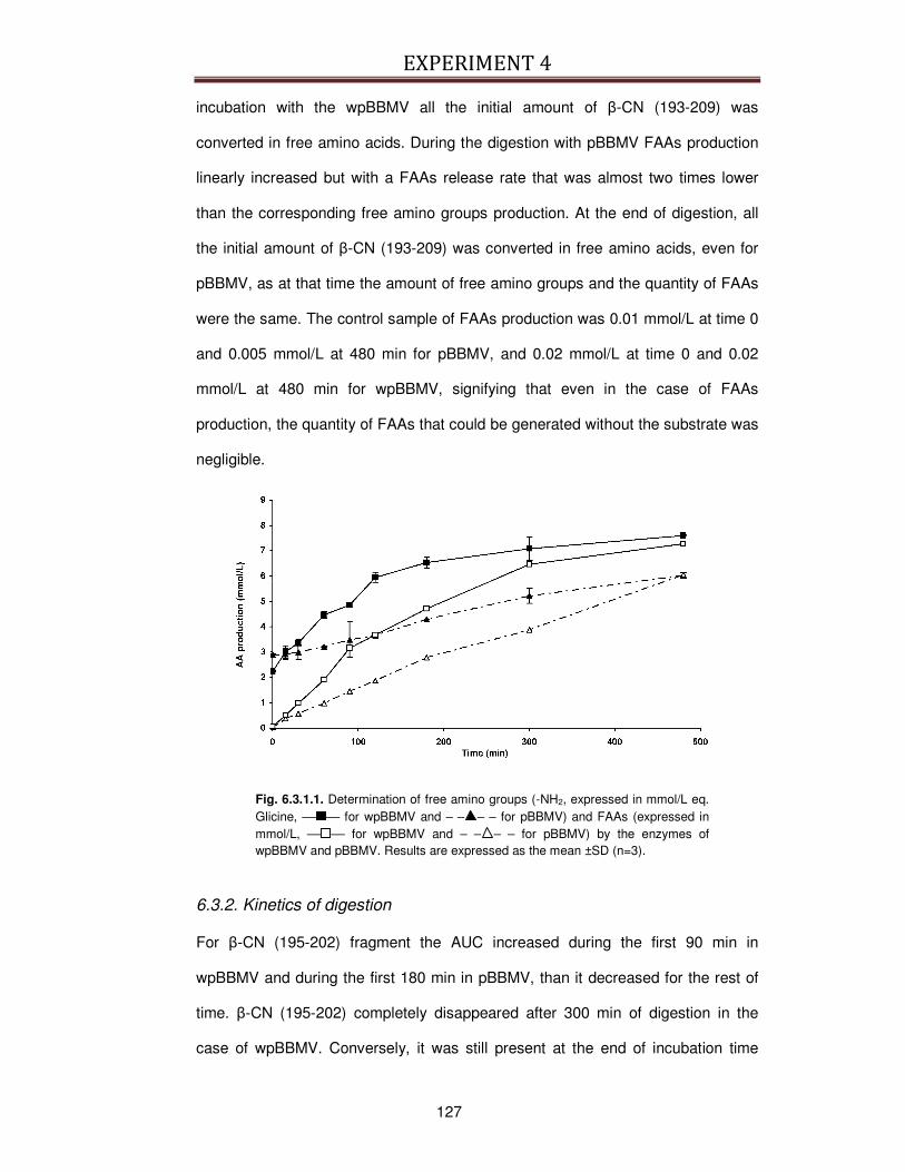
EXPERIMENT 4
127
incubation with the wpBBMV all the initial amount of β-CN (193-209) was
converted in free amino acids. During the digestion with pBBMV FAAs production
linearly increased but with a FAAs release rate that was almost two times lower
than the corresponding free amino groups production. At the end of digestion, all
the initial amount of β-CN (193-209) was converted in free amino acids, even for
pBBMV, as at that time the amount of free amino groups and the quantity of FAAs
were the same. The control sample of FAAs production was 0.01 mmol/L at time 0
and 0.005 mmol/L at 480 min for pBBMV, and 0.02 mmol/L at time 0 and 0.02
mmol/L at 480 min for wpBBMV, signifying that even in the case of FAAs
production, the quantity of FAAs that could be generated without the substrate was
negligible.
Fig. 6.3.1.1. Determination of free amino groups (-NH2, expressed in mmol/L eq. Glicine, –– –– for wpBBMV and – – – – for pBBMV) and FAAs (expressed in mmol/L, –– –– for wpBBMV and – – – – for pBBMV) by the enzymes of wpBBMV and pBBMV. Results are expressed as the mean ±SD (n=3).
6.3.2. Kinetics of digestion
For β-CN (195-202) fragment the AUC increased during the first 90 min in
wpBBMV and during the first 180 min in pBBMV, than it decreased for the rest of
time. β-CN (195-202) completely disappeared after 300 min of digestion in the
case of wpBBMV. Conversely, it was still present at the end of incubation time
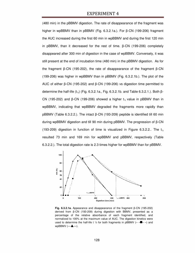
EXPERIMENT 4
128
(480 min) in the pBBMV digestion. The rate of disappearance of the fragment was
higher in wpBBMV than in pBBMV (Fig. 6.3.2.1a.). For β-CN (199-206) fragment
the AUC increased during the first 60 min in wpBBMV and during the first 120 min
in pBBMV, than it decreased for the rest of time. β-CN (199-206) completely
disappeared after 300 min of digestion in the case of wpBBMV. Conversely, it was
still present at the end of incubation time (480 min) in the pBBMV digestion. As for
the fragment β-CN (195-202), the rate of disappearance of the fragment β-CN
(199-206) was higher in wpBBMV than in pBBMV (Fig. 6.3.2.1b.). The plot of the
AUC of either β-CN (195-202) and β-CN (199-206) vs digestion time permitted to
determine the half-life (t½) (Fig. 6.3.2.1a., Fig. 6.3.2.1b. and Table 6.3.2.1.). Both β-
CN (195-202) and β-CN (199-206) showed a higher t½ value in pBBMV than in
wpBBMV, indicating that wpBBMV degraded the fragments more rapidly than
pBBMV (Table 6.3.2.2.). The intact β-CN (193-209) peptide is identified till 60 min
during wpBBMV digestion and till 90 min during pBBMV. The progression of β-CN
(193-209) digestion in function of time is visualized in Figure 6.3.2.2.. The t½
resulted 73 min and 169 min for wpBBMV and pBBMV, respectively (Table
6.3.2.2.). The total digestion rate is 2.3 times higher for wpBBMV than for pBBMV.
Fig. 6.3.2.1a. Appearance and disappearance of the fragment β-CN (195-202) derived from β-CN (193-209) during digestion with BBMV, presented as a percentage of the relative absorbance of each fragment identified, and normalized to 100% at the maximum value of AUC. The digestion kinetics were used to determine the half-life t ½ for both fragments in pBBMV (– – – –) and wpBBMV (–– ––).
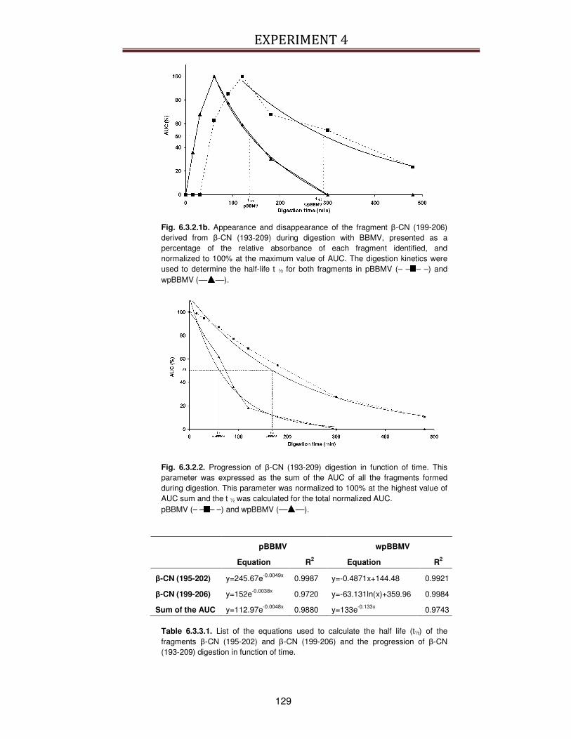
EXPERIMENT 4
129
Fig. 6.3.2.1b. Appearance and disappearance of the fragment β-CN (199-206) derived from β-CN (193-209) during digestion with BBMV, presented as a percentage of the relative absorbance of each fragment identified, and normalized to 100% at the maximum value of AUC. The digestion kinetics were used to determine the half-life t ½ for both fragments in pBBMV (– – – –) and wpBBMV (–– ––).
Fig. 6.3.2.2. Progression of β-CN (193-209) digestion in function of time. This parameter was expressed as the sum of the AUC of all the fragments formed during digestion. This parameter was normalized to 100% at the highest value of AUC sum and the t ½ was calculated for the total normalized AUC. pBBMV (– – – –) and wpBBMV (–– ––).
pBBMV wpBBMV
Equation R2 Equation R2
β-CN (195-202) y=245.67e-0.0049x 0.9987 y=-0.4871x+144.48 0.9921
β-CN (199-206) y=152e-0.0038x 0.9720 y=-63.131ln(x)+359.96 0.9984
Sum of the AUC y=112.97e-0.0048x 0.9880 y=133e-0.133x 0.9743
Table 6.3.3.1. List of the equations used to calculate the half life (t½) of the fragments β-CN (195-202) and β-CN (199-206) and the progression of β-CN (193-209) digestion in function of time.
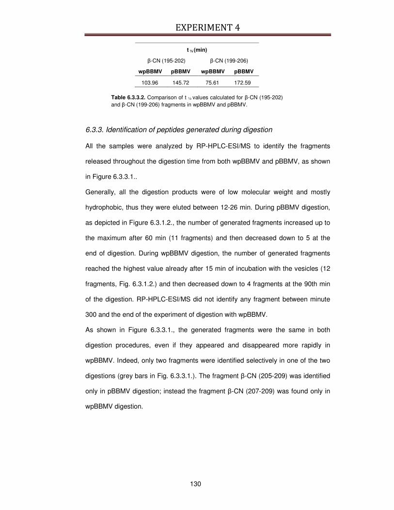
EXPERIMENT 4
130
t ½ (min)
β-CN (195-202) β-CN (199-206)
wpBBMV pBBMV wpBBMV pBBMV
103.96 145.72 75.61 172.59
Table 6.3.3.2. Comparison of t ½ values calculated for β-CN (195-202) and β-CN (199-206) fragments in wpBBMV and pBBMV.
6.3.3. Identification of peptides generated during digestion
All the samples were analyzed by RP-HPLC-ESI/MS to identify the fragments
released throughout the digestion time from both wpBBMV and pBBMV, as shown
in Figure 6.3.3.1..
Generally, all the digestion products were of low molecular weight and mostly
hydrophobic, thus they were eluted between 12-26 min. During pBBMV digestion,
as depicted in Figure 6.3.1.2., the number of generated fragments increased up to
the maximum after 60 min (11 fragments) and then decreased down to 5 at the
end of digestion. During wpBBMV digestion, the number of generated fragments
reached the highest value already after 15 min of incubation with the vesicles (12
fragments, Fig. 6.3.1.2.) and then decreased down to 4 fragments at the 90th min
of the digestion. RP-HPLC-ESI/MS did not identify any fragment between minute
300 and the end of the experiment of digestion with wpBBMV.
As shown in Figure 6.3.3.1., the generated fragments were the same in both
digestion procedures, even if they appeared and disappeared more rapidly in
wpBBMV. Indeed, only two fragments were identified selectively in one of the two
digestions (grey bars in Fig. 6.3.3.1.). The fragment β-CN (205-209) was identified
only in pBBMV digestion; instead the fragment β-CN (207-209) was found only in
wpBBMV digestion.
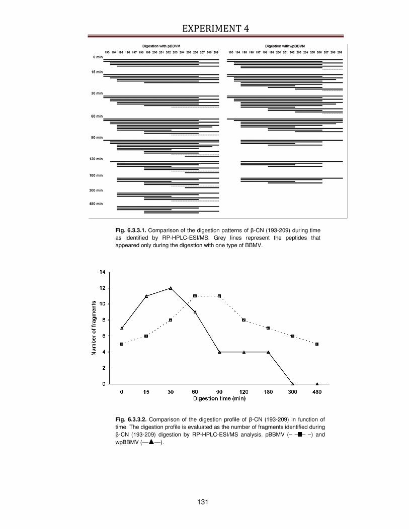
EXPERIMENT 4
131
Fig. 6.3.3.1. Comparison of the digestion patterns of β-CN (193-209) during time as identified by RP-HPLC-ESI/MS. Grey lines represent the peptides that appeared only during the digestion with one type of BBMV.
Fig. 6.3.3.2. Comparison of the digestion profile of β-CN (193-209) in function of time. The digestion profile is evaluated as the number of fragments identified during β-CN (193-209) digestion by RP-HPLC-ESI/MS analysis. pBBMV (– – – –) and wpBBMV (–– ––).
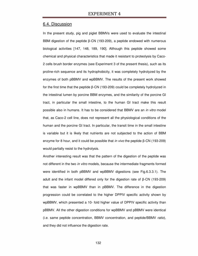
EXPERIMENT 4
132
6.4. Discussion
In the present study, pig and piglet BBMVs were used to evaluate the intestinal
BBM digestion of the peptide β-CN (193-209), a peptide endowed with numerous
biological activities [147, 148, 189, 190]. Although this peptide showed some
chemical and physical characteristics that made it resistant to proteolysis by Caco-
2 cells brush border enzymes (see Experiment 3 of the present thesis), such as its
proline-rich sequence and its hydrophobicity, it was completely hydrolyzed by the
enzymes of both pBBMV and wpBBMV. The results of the present work showed
for the first time that the peptide β-CN (193-209) could be completely hydrolyzed in
the intestinal lumen by porcine BBM enzymes, and the similarity of the porcine GI
tract, in particular the small intestine, to the human GI tract make this result
possible also in humans. It has to be considered that BBMV are an in vitro model
that, as Caco-2 cell line, does not represent all the physiological conditions of the
human and the porcine GI tract. In particular, the transit time in the small intestine
is variable but it is likely that nutrients are not subjected to the action of BBM
enzyme for 8 hour, and it could be possible that in vivo the peptide β-CN (193-209)
would partially resist to the hydrolysis.
Another interesting result was that the pattern of the digestion of the peptide was
not different in the two in vitro models, because the intermediate fragments formed
were identified in both pBBMV and wpBBMV digestions (see Fig.6.3.3.1). The
adult and the infant model differed only for the digestion rate of β-CN (193-209)
that was faster in wpBBMV than in pBBMV. The difference in the digestion
progression could be correlated to the higher DPPIV specific activity shown by
wpBBMV, which presented a 10- fold higher value of DPPIV specific activity than
pBBMV. All the other digestion conditions for wpBBMV and pBBMV were identical
(i.e. same peptide concentration, BBMV concentration, and peptide/BBMV ratio),
and they did not influence the digestion rate.
EXPERIMENT 4
133
The identification of fragments generated by RP-HPLC-ESI/MS allowed making
some hypothesis on the enzymes that intervened during the digestions. In both
cases, the bond Tyr193-Gln194 at the N-terminus could be the first to be cleaved by
an exopeptidase, subsequently followed by the bond Gln194-Glu195, always by the
action of an exopeptidase. At the C-terminus, the action of exopeptidases could
have been less marked than at the N-terminus because of the cleavage of the
bond Pro206-Ile207 by an endopeptidase that released the fragment β-CN (207-209).
The endopeptidases could be responsible also of the cleavage of the bonds
Leu198-Gly199 and Arg202-Gly203. In particular, the endopeptidases generated the
fragment β-CN (195-202) that resisted to the proteolytic action of vesicles enzyme
up to the end of digestion for pBBMV and up to 180 min for wpBBMV.
The assessment of digestion by monitoring the free amino groups (-NH2) and
FAAs amount in function of time allowed the comparison of the endopeptidases
activity to the activity of exopeptidases (Fig. 6.3.1.1.). At the end of the digestion
time, the amount of free amino groups overlapped the quantity of FAAs in both
wpBBMV and pBBMV, demonstrating that all the generated fragments (quantified
as -NH2 groups) were completely hydrolyzed to free amino acids (quantified as
FAAs). Thus, the exopeptidases activity mainly contributed to the digestion of the
peptide β-CN (193-209).
It is of note that 8 hours of digestion is not necessarily representative of a digestion
process in vivo [360], because the ratio between the concentration of the peptide/
and the concentration of BBMV has been selected to better monitor the digestion
kinetics and understand the mechanisms and possible differences in the pattern of
digestion between the adult (pBBMV) and the infant models (wpBBMV).
The peptide β-CN (193-209) has to remain intact in the intestine to express its
biological activity in vivo, while in the present work, the peptide was completely
digested by BBMV enzymes. The BBMV represents an in vitro model of the BBM
of the enterocytes, enriched in proteolytic enzymes in comparison to the intestinal

EXPERIMENT 4
134
enterocytes. For this reason, it could be possible that in physiological conditions
and transit time, β-CN (193-209) peptide would resist to complete hydrolysis.
A better knowledge of the digestion of bioactive peptides is needed to understand
their in vivo stability in the intestinal tract, and the mechanisms of interaction with
the intestinal mucosa [360]. With this purpose, the present study on the in vitro
simulated BBM digestion of the immunomodulatory peptide β-CN (193-209)
permitted to clarify the enzyme category that mostly intervene in the digestion of
this peptide and the most probable cleavage sites in its sequence.
In conclusion, the present work demonstrated that BBMV completely degraded β-
CN (193-209) peptide and that the most involved enzymes are exopeptidases. In
addition the present work confirmed the usefulness of BBMV as a tool to
understand the mechanisms that determine the digestion profile of the bioactive β-
CN (193-209). Further work is needed to integrate all the data regarding the
stability of this peptide and other similar bioactive peptides in different in vitro
gastrointestinal models to identify the most important characteristics that contribute
to bioactive peptide bioavailability.
6.5. Take-home message
The present study demonstrated that the immunomodulatory peptide β-CN (193-
209) obtained from bovine β-casein is completely digested by the BBM enzymes of
porcine BBMV, an in vitro model for the intestinal epithelium. This is an important
result that integrates the data obtained on the digestion of the peptide achieved
from the Experiment 3, using Caco-2 monolayers. However, these results should
be completed with other in vitro models that obviate the limitations of Caco-2 cell
line and BBMV, and that better represent the physiological conditions of the
digestion process. In this sense, it can be hypothesized that primary cell cultures

EXPERIMENT 4
135
from porcine gut mucosa would give helpful data because they are more similar in
their phenotype to the mature enterocyte.
GENERAL DISCUSSION
137
GENERAL DISCUSSION
7.1. Studies on the digestion and absorption of bioactive peptides
The Experiments 3 and 4 explored how the immunomodulatory peptide β-CN (193-
209) generated from β-casein is digested and absorbed in two models for the
human GI tract. In particular, the important result of this work is that this peptide
could resist to hydrolysis by intestinal proteases and peptidases and a part of the
starting amount given to the cells could by absorbed intact by the cell monolayer.
However, a lot of work should be done to better understand the mechanisms that
permitted the absorption, and the enzymes that primarily intervene in the digestion
of the model peptide β-CN (193-209) should be identified.
At the moment many models for human digestion and absorption of different
nutrients in the GI tract are available (see Paragraphs 2.4. and 2.5.), but none of
them is specifically made for the evaluation absorption and digestion of bioactive
peptides. The main characteristics that should be taken into consideration in the
realization of a specific model should be the role of the proteases and peptidases of
the GI tract in liberating bioactive peptides that are not already present in a free and
active form in food matrices and in hydrolyzing some other bioactive peptides
already present in the food.
It may be hypothesized that the already established dynamic models or the
computer-controlled system that at present better represent the human GI complex
physiology should be first modified for the integrated study of digestion and
absorption and also characterized for the most important enzymes involved in the
generation and stability of bioactive peptides.
Some useful implementations should be directed also in the field of in vitro models
based on cell lines. A better prediction of absorption could be gained, if the apical
pH is 5.5–6.5 and this can be achieved without compromising the integrity of Caco-2
GENERAL DISCUSSION
138
cell monolayers, as demonstrated by Palm and colleagues [289] and Yamashita and
colleagues [290]. The change in pH has been evaluated in n-in-one permeability
studies for passively permeated drugs [291] and the authors found that Caco-2 cells
better mimicked the in vivo conditions and gave more reliable information about the
absorption of drugs across the enterocytic membrane, so it could be hypothesized
that the same more reliable results could be also obtained in the case of the
evaluation of bioactive peptide absorption.
Complementary information on the absorption of bioactive peptides could be gained
from cell lines other than Caco-2. It is well known [292] that permeabilities of
compounds that are transported via carrier-mediated absorption are lower in the
Caco-2 cell system as compared to the human small intestine, probably reflecting
the colonic origin of this cell line. In recent years several mucus-producing goblet
cell sublines have been established from human intestinal HT29 cells, as HT29-MTX
[293-295], a cell population that consists exclusively of differentiated, gastric-like
mucus secreting, goblet-type cells that retain their differentiated phenotype after
reversion to a methotrexate (MTX)-free medium and they also can be grown in
monolayers. At present these cells lines are mostly used for drug absorption studies
but it cannot be excluded a potential application in bioactive peptide absorption
evaluation. A possible implementation of the Caco-2 cell line model could be the use
of cell lines transfected with a specific oligopeptide transporter for the evaluation of
the structural features required for interaction and transport. In addition, helpful
information could be acquired using primary cell cultures isolated from the intestine
mucosa, an expanding research area, as demonstrated by the increasing number of
reports focusing in particular on bovine intestine cell culture [371-373].

GENERAL DISCUSSION
139
Some studies on the digestion and absorption of bioactive peptides derived from
milk-proteins, with special attention to ACE-inhibitory ones, have already
demonstrated that some bioactive tripeptides could be absorbed. For example, VPP
was detected in the abdominal aorta of spontaneous hypertensive rats (SHR) 6
hours after its administration in sour milk, which strongly suggests that it is
transepithelially transported [260]; more recently the absorption was observed also
in humans [6]. Paracellular transport, through the intercellular junctions, was
suggested as the main mechanism, since the transport via the short-peptide carrier,
PepT1, led to a quick hydrolysis of the internalized peptide [186]. In the case of
larger sequences, the susceptibility to brush border peptidases is the primary factor
that decides the transport rate [244]. For example, the heptapeptide lactokinins
(ALPMHIR) was transported intact, although in concentrations too low to exert an
ACE-inhibitory activity, which suggests cleavage by aminopeptidases [261].
The Experiment 3 of the present thesis gave some insight on the absorption of long
bioactive peptides. In fact, in the case of the peptide β-CN (193-209), the result
obtained leaded to the hypothesis that its transport could be mainly mediated by
transcytosis, even if a role of the paracellular transport could not be completely
excluded. No data were obtained about a possible energy-dependent transporter for
long peptides.
More research is needed in this respect, with the effort being concentrated in
elucidating the pharmacokinetics and the distribution profile of milk-derived bioactive
peptides in the different tissues.
7.2. The evaluation of the immunomodulatory activity of bioactive
peptides
From the present thesis, in particular from Experiments 1 and 2, some questions
have risen about the best method that would permit a reliable evaluation of the
GENERAL DISCUSSION
140
effective activity of bioactive peptides and milk-derived products with a potential
immunomodulatory action. Indeed, the immunomodulatory effects of yogurt (i. e.
against cancer) have been studied, mostly using animal models [72, 78, 79, 97-103].
Conversely, few human studies on the immunostimulatory effects of yogurt and
immunomodulatory peptides have been conducted [7]. Although the results of these
studies mostly support the notion that yogurt has immunostimulatory effects, poor
study design, lack of appropriate controls, and short duration of most of the studies
limit the value of the conclusions that can be drawn from them. Most early animal
and human studies included too few subjects in each group, and most of them did
not include statistical analysis. Even in animal studies, the majority used short-term
feeding protocols, which might induce a transient adjuvant effect rather than a long-
term stimulation of the immune response. Furthermore, most studies investigated
the effect of intravenous or intraperitoneal administration or in vitro application of
yogurt and immunomodulatory peptides on different variables of the immune
response but, as immunomodulatory peptides are consumed orally and they may be
altered in the GI tract, the results of these studies may not reflect what would be
found if the yogurt had been consumed orally.
The main problem however is that all the studies investigated the effects of
immunomodulatory peptides or yogurt on in vitro indexes of the immune response
(i.e. lymphocytes proliferation by DNA or protein synthesis, or antibody production,
or cytotoxicity ability) and these parameters could not represent the complexity of
the variables of the whole immune system in vivo. As a consequence, the
preliminary result obtained on the immunomodulatory activity of the peptide YGG
and on the milk fermented by L. delb. bulgaricus LA2 should be confirm in an in vivo
model, taking into account the modification on the bioactive peptide operated by the
GI tract and considering also the fact that the gut-associated immune system is
increasingly being recognized as playing an important role in host defense. In fact,
the M-cells of the Peyer’s Patches dispersed in the intestinal mucosa may contribute
GENERAL DISCUSSION
141
to the translocation of intact bioactive peptides across the intestinal epithelium, thus
increasing the possibility of these peptides to act on the different subpopulations of
intestinal cells Otani, 1995 111 /id. This aspect of the immune response is
particularly relevant to determining the beneficial effects of bioactive peptides
because their systemic effects may depend on the interaction of the peptides with
the immune cells of the gut. However, the interactions between immunomodulatory
peptides and the gut-associated immune system has been scarcely explored [51,
52, 132, 162] and thus expanding the knowledge in this field would be of extreme
importance.
7.3. Future perspectives on the production of dairy food with ACE-
inhibitory and immunomodulatory properties
Experiment 1 gave some preliminary and encouraging results about the possibility to
generate fermented milk with ACE-inhibitory activity by a bacterial strain belonging
to E. faecalis species. This result confirms the fact that bioactive peptides and milk-
derived products with antihypertensive or immunomodulatory properties can be
produced in different ways but fermentation with LAB is the preferred one.
Expanding the knowledge about the proteolytic systems of interesting LAB, and their
activity under various conditions, more specifically belonging to E. Faecalis species,
could be a relevant step to improve the amount and the stability of ACE-inhibitory
peptides in the dairy products [125]. In addition, further progress in this area might
be obtained through genetic engineering, to provide the most suited strain with the
desired proteolytic capacity, and also from studies regarding the interaction between
strains in environments as those prevailing in fermented milks and cheeses [125].
Moreover, regardless the source containing bioactive peptides and the associated
bioactivity, it is important that bioactive peptides must be stable during the final
processing, packaging and storage. Furthermore, the hydrolysate should have well-
GENERAL DISCUSSION
142
defined technological functionalities not to impart the required functionality of the
carrier food [125].
Hence, more information must be acquired on the influence of food processing,
preparation and preservation on the bioactivity of bioactive peptides.
CONCLUSIONS
143
CONCLUSIONS
The review of literature and the results obtained in the present thesis suggest that
food microorganisms, isolated from food matrices, in particular of bacterial origin, act
on the nutrients contained in the food. These microorganisms could thus generate
functional foods enriched in specific components able to influence important
physiological processes of the human body, as blood pressure or immune response.
In this view, the present work explored the possibility to use E. faecalis TH563 to
produce fermented milk with ACE-inhibitory activity and L. delb. bulgaricus LA2 to
obtain fermented milk with immunomodulatory activity, even if it would be necessary
to evaluate E. faecalis TH563 for safety aspects.
As a consequence, there is an increasing need to select the microorganism present
in food matrices for their ability to produce functional food enriched in specific
bioactivities on large scale. More research is thus needed to characterize the
microorganisms and the associated bioactivities and to develop new methods
permitting the unambiguous quantification of the bioactivity in the foodstuff and the
identification of the food components responsible of such bioactivity. For example, it
would be interesting to identify the presence of the peptides β-CN (193-209) and
YGG in milk fermented by L. delb. bulgaricus LA2 to acquire better knowledge about
the mechanisms determining the associated immunomodulatory activity.
With this purpose, the Experiments 1 and 2 have been realized. They aimed to
study the immunomodulatory activity of the milk fermented by two bacterial strains
frequently found in dairy products of the North-East of Italy and to clarify the
mechanism of action of a milk-derived peptide with already documented
immunomodulatory activity on lymphocytes, considered as a model peptide, as the
peptide YGG derived from α-lactalbumin. The present work demonstrated that YGG
modulated bovine lymphocyte proliferation and that this effect is dependent upon
serum concentration and on the presence of lymphocyte activators, such as
CONCLUSIONS
144
concanavalin A, in the culture medium. Nevertheless, it has been observed that the
YGG effects on lymphocyte proliferation did not seem to be mediated by a
modulation of the RNA expression of IL2 and INFγ, two important cytokines involved
in lymphocytes activation and proliferation. The obtained results, together with the
Paragraph 2.3.2., demonstrate that the in vitro methods manifest some limitations in
the characterization of immunomodulatory bioactivity and that an exhaustive view of
the action of immunomodulatory peptides could be achieved only by a multi-view
approach that should take into account the complexity of the interactions between
the bioactive peptide and the different components of the immune system in vivo. In
fact, the Experiment 1 and the Paragraph 7.1. in the General Discussion section
evidence the lack of knowledge about the interaction of the immunomodulatory
peptides derived from food and the immune system dispersed along the GI tract (as
GALT, Peyer’s Patches, antigen-presenting cells) that could represent a potential
target of immunomodulatory peptides, even before to be absorbed at gut level and
circulate in the body.
At the moment the interactions between food-derived peptides and the gut-
associated immune system have been explored to elucidate the mechanisms
underlying allergies but it would be interesting to apply the same approach to
evaluate the bioactivities, considering both allergies and bioactivities as properties
that could be displayed by peptides.
The present thesis focused also on the physiology of absorption of bioactive
peptides and demonstrated for the first time that a long hydrophobic bioactive
peptide crossed intact a Caco-2 cell monolayer, a well recognized in vitro model for
the intestinal epithelium. In fact, the milk-derived immunomodulatory peptide β-CN
(193-209) was demonstrated to be resistant to the digestion of gastrointestinal
peptidases and to pass intact across Caco-2 cells. In addition, the digestion profile
of this peptide has been studied in brush border membrane vesicles.
CONCLUSIONS
145
This interesting result permits to suggest that even large peptides could be
absorbed in small quantities and that it cannot be excluded that at these
concentrations the peptide β-CN (193-209) could interact with the gut-associated
immune system, as explained before.
As a consequence, the assessment of the digestion profile and of the mechanism of
absorption of β-CN (193-209) could be considered as model studies for the
evaluation of the bioavailability of bioactive peptides, such as YG peptide. In fact, it
would be helpful to examine the bioavailability of this bioactive peptide, checking for
the resistance to gastrointestinal and serum peptidases. For example, it would be
interesting to identify the presence of the peptides β-CN (193-209) and YGG in milk
fermented by L. delb. bulgaricus LA2 to acquire better knowledge about the
mechanisms determining the associated bioactivity.
In conclusion, new questions have arisen on the area of bioactive peptides that
could constitute the objective of further research studies in the future.
ACKNOWLEDGEMENTS
147
ACKNOWLEDGEMENTS
I wish to acknowledge the many people both in Italy and in France that have given
me their support during the development of this thesis.
I am very grateful to my supervisors Prof. Alessandro Negro and Prof. Gianfranco
Gabai for their assistance in completing this project.
I am also particularly grateful to Dr. Joelle Leonil, Dr Rachel Boutrou and Dr. Didier
Dupont of the INRA UMR 1253 “STLO” at Rennes that gave me the possibility to
spend a part of my PhD project in the INRA laboratories, guiding me during my “PhD
experience” and teaching me much more than laboratory techniques and
interpretation of results.
I would also like to acknowledge all the academics, staff and students from the
laboratories of “Dipartimento di Scienze Sperimentali Veterinarie” (Università degli
Studi di Padova). Special thanks to Laura Dadalt, Tommaso Brogin and Giovanni
Caporale for helping with me during this project. Extended thanks to Lisa
Maccatrozzo who guided me in the Real-Time PCR analyses.
Moreover, I would also like to acknowledge all the people of the UMR 1253 “Science
et Technologie du Lait et de l’Œuf”, in particular Daniel Mollé et Julien Jardin for all
the mass spectrometry analyses and for their patient help in the interpretation of the
MS profiles.
A special thank also to the staff of “Azienda Sperimentale Veterinaria L. Toniolo”
(Università degli Studi di Padova), that collected the blood samples for the isolation
of bovine lymphocytes and a very special thank to Christian Andrighetto and
Angiolella Lombardi of the Institute of Veneto Agricoltura of Thiene, who kindly
supplied the bacterial strains and introduced me in the world of dairy
microorganisms.
Last but obviously not the least, I would like to thank Riccardo who always
supported me during this long and difficult experience, encouraging and helping me

ACKNOWLEDGEMENTS
148
to find a meaning in all the parts of this PhD project, when it was not so immediate
for me.

WEB REFERENCES
149
WEB REFERENCES http://en.wikipedia.org/wiki//File:Renin-angiotensin-aldosterone_system.png
http://en.wikipedia.org/wiki/Immune_system
http://en.wikipedia.org/wiki/T_cell
http://en.wikipedia.org/wiki/Cytokine
http://kvhs.nbed.nb.ca/gallant/biology/small_intestine.jpg
http://microvet.arizona.edu/courses/MIC419/Tutorials/cytokines.html
http://www.foodsci.uoguelph.ca/dairyedu/home.html
http://www.uta.edu/chagas/html/biolImS1.html
http://www.wzw.tum.de/nutrition/index.php?id=31.
http://www.warneronline.com/product_info.cfm?name=Introduction to Ussing
Chamber and System from Warner&id=1401
www.cancer.gov/cancertopics/understandingcancer/immunesystem/
www.acm.uiuc.edu/sigbio/project/digestive/middle/stomach2.jpg
www.colorado.edo/intphys/class/IPHY3430-200/image/21-25.jpg
www.yoursurgery.com/procedures/intussussception/images/SmBowelAnat.jpg
www.fi.cnr.it/r&f/n4/images/spadoni.jpg

REFERENCES
150
REFERENCES
[1] Cross ML, Gill HS. Immunomodulatory properties of milk. Br J Nutr 2000; 84 Suppl 1: S81-S89.
[2] Newby TJ, Stokes CR, Bourne FJ. Immunological activities of milk. Vet Immunol Immunopathol 1982; 3: 67-94.
[3] Tamime AY. Fermented milks: a historical food with modern applications--a review. Eur J Clin Nutr 2002; 56 Suppl 4: S2-S15.
[4] Rachid M, Matar C, Duarte J, Perdigon G. Effect of milk fermented with a Lactobacillus helveticus R389(+) proteolytic strain on the immune system and on the growth of 471 breast cancer cells in mice. FEMS Immunol Med Microbiol 2006; 47: 242-253.
[5] Seppo L, Jauhiainen T, Poussa T, Korpela R. A fermented milk high in bioactive peptides has a blood pressure-lowering effect in hypertensive subjects. Am J Clin Nutr 2003; 77: 326-330.
[6] Foltz M, Meynen EE, Bianco V, van Platerink C, Koning TMMG, Kloek J. Angiotensin converting enzyme inhibitory peptides from a lactotripeptide-enriched milk beverage are absorbed intact into the circulation. J Nutr 2007; 137: 953-958.
[7] Parra MD, Martinez de Morentin BE, Cobo JM, Mateos A, Martinez JA. Daily ingestion of fermented milk containing Lactobacillus casei DN114001 improves innate-defense capacity in healthy middle-aged people. J Physiol Biochem 2004; 60: 85-91.
[8] Kawase M, Hashimoto H, Hosoda M, Morita H, Hosono A. Effect of administration of fermented milk containing whey protein concentrate to rats and healthy men on serum lipids and blood pressure. J Dairy Sci 2000; 83: 255-263.
[9] Moller NP, Scholz-Ahrens KE, Roos N, Schrezenmeir J. Bioactive peptides and proteins from foods: indication for health effects. Eur J Nutr 2008; 47: 171-182.
[10] Hata Y, Yamamoto M, Ohni M, Nakajima K, Nakamura Y, Takano T. A placebo-controlled study of the effect of sour milk on blood pressure in hypertensive subjects. Am J Clin Nutr 1996; 64: 767-771.
[11] Mizushima S, Ohshige K, Watanabe J, Kimura M, Kadowaki T, Nakamura Y et al. Randomized controlled trial of sour milk on blood pressure in borderline hypertensive men. Am J Hypertens 2004; 17: 701-706.
[12] Nakamura Y, Yamamoto N, Sakai K, Takano T. Antihypertensive effect of sour milk and peptides isolated from it that are inhibitors to Angiotensin I-converting enzyme. J Dairy Sci 1995; 78: 1253-1257.
REFERENCES
151
[13] Tellez A, Corredig M, Brovko LY, Griffiths MW. Characterization of immune-active peptides obtained from milk fermented by Lactobacillus helveticus. J Dairy Res 2010; Jan 18: 1-8.
[14] Hata I, Ueda J., Otani H. Immunostimulatory action of a commercially available casein phosphopeptide preparation, CPP-III, in cell cultures. Milchwissenschaften 1999; 54: 3-7.
[15] Laffineur E, Genetet N, Leonil J. Immunomodulatory activity of beta-casein permeate medium fermented by lactic acid bacteria. J Dairy Sci 1996; 79: 2112-2120.
[16] Clare DA, Catignani GL, Swaisgood HE. Biodefense properties of milk: the role of antimicrobial proteins and peptides. Curr Pharm Des 2003; 9: 1239-1255.
[17] Tuomilehto J, Lindstrom J, Hyyrynen J, Korpela R, Karhunen ML, Mikkola L et al. Effect of ingesting sour milk fermented using Lactobacillus helveticus bacteria producing tripeptides on blood pressure in subjects with mild hypertension. J Hum Hypertens 2004; 18: 795-802.
[18] Schrezenmeir J, Korhonen H, Williams C, Gill HS, Shah N. Beneficial natural bioactive substances in milk and colostrum - Occurrence, biochemical and technological characteristics of bioactive substances - Physiological effects and potential health benefits - Foreword. Br J Nutr 2000; 84: S1.
[19] Korhonen H, Pihlanto A. Food-derived bioactive peptides--opportunities for designing future foods. Curr Pharm Des 2003; 9: 1297-1308.
[20] Clare DA, Swaisgood HE. Bioactive milk peptides: a prospectus. J Dairy Sci 2000; 83: 1187-1195.
[21] Majumder K, Wu J. Angiotensin I converting enzyme inhibitory peptides from simulated in vitro gastrointestinal digestion of cooked eggs. J Agric Food Chem 2009; 57: 471-477.
[22] Bauchart C, Morzel M, Chambon C, Mirand PP, Reynes C, Buffiere C et al. Peptides reproducibly released by in vivo digestion of beef meat and trout flesh in pigs. Br J Nutr 2007; 98: 1187-1195.
[23] Leadbeater L, Ward FB. Analysis of tryptic digests of bovine beta-casein by reversed-phase high-performance liquid chromatography. J Chromatogr 1987; 397: 435-443.
[24] Lemieux L, Amiot J. High-performance liquid chromatography of casein hydrolysates phosphorylated and dephosphorylated. I. Peptide mapping. J Chromatogr 1990; 519: 299-321.
[25] Svedberg J, de HJ, Leimenstoll G, Paul F, Teschemacher H. Demonstration of beta-casomorphin immunoreactive materials in in vitro digests of bovine milk and in small intestine contents after bovine milk ingestion in adult humans. Peptides 1985; 6: 825-830.
[26] Miquel E, Gomez JA, Alegria A, Barbera R, Farre R, Recio I. Identification of casein phosphopeptides released after simulated digestion of milk-based infant formulas. J Agric Food Chem 2005; 53: 3426-3433.

REFERENCES
152
[27] Matar C, Amiot J, Savoie L, Goulet J. The effect of milk fermentation by Lactobacillus helveticus on the release of peptides during in vitro digestion. J Dairy Sci 1996; 79: 971-979.
[28] Christensen JE, Dudley EG, Pederson JA, Steele JL. Peptidases and amino acid catabolism in lactic acid bacteria. Antonie Van Leeuwenhoek 1999; 76: 217-246.
[29] Gobbetti M, Ferranti P, Smacchi E, Goffredi F, Addeo F. Production of angiotensin-I-converting-enzyme-inhibitory peptides in fermented milks started by Lactobacillus delbrueckii subsp. bulgaricus SS1 and Lactococcus lactis subsp. cremoris FT4. Appl Environ Microbiol 2000; 66: 3898-3904.
[30] Pihlanto-Leppala A, Rokka T, Korhonen H. Angiotensin I converting enzyme inhibitory peptides derived from bovine milk proteins. Int Dairy J 1998; 8: 325-331.
[31] Gill HS, Doull F, Rutherfurd KJ, Cross ML. Immunoregulatory peptides in bovine milk. Br J Nutr 2000; 84 Suppl 1: S111-S117.
[32] Gobbetti M, Minervini F, Rizzello CG. Angiotensin I-converting-enzyme-inhibitory and antimicrobial bioactive peptides. Int J Dairy Technol 2004; 57: 173-188.
[33] FitzGerald RJ, Murray BA, Walsh DJ. Hypotensive peptides from milk proteins. J Nutr 2004; 134: 980S-988S.
[34] Meisel H, FitzGerald RJ. Biofunctional peptides from milk proteins: mineral binding and cytomodulatory effects. Curr Pharm Des 2003; 9: 1289-1295.
[35] Yamamoto N, Ejiri M, Mizuno S. Biogenic peptides and their potential use. Curr Pharm Des 2003; 9: 1345-1355.
[36] Tauzin J, Miclo L, Gaillard JL. Angiotensin-I-converting enzyme inhibitory peptides from tryptic hydrolysate of bovine alphaS2-casein. FEBS Lett 2002; 531: 369-374.
[37] Manso MA, Lopez-Fandino R. Angiotensin I converting enzyme-inhibitory activity of bovine, ovine, and caprine kappa-casein macropeptides and their tryptic hydrolysates. J Food Prot 2003; 66: 1686-1692.
[38] Kilara A, Panyam D. Peptides from milk proteins and their properties. Crit Rev Food Sci Nutr 2003; 43: 607-633.
[39] Yamamoto N, Akino A, Takano T. Antihypertensive effect of the peptides derived from casein by an extracellular proteinase from Lactobacillus helveticus CP790. J Dairy Sci 1994; 77: 917-922.
[40] Maeno M, Yamamoto N, Takano T. Identification of an antihypertensive peptide from casein hydrolysate produced by a proteinase from Lactobacillus helveticus CP790. J Dairy Sci 1996; 79: 1316-1321.
[41] Mizuno S, Nishimura S, Matsuura K, Gotou T, Yamamoto N. Release of short and proline-rich antihypertensive peptides from casein hydrolysate with an Aspergillus oryzae protease. J Dairy Sci 2004; 87: 3183-3188.

REFERENCES
153
[42] Teschemacher H, Koch G, Brantl V. Milk protein-derived opioid receptor ligands. Biopolymers 1997; 43: 99-117.
[43] Bal dit SC, Drouet L, Pignaud G, Chevallier C, Caen J, Fiat AM et al. Effect of kappa-casein split peptides on platelet aggregation and on thrombus formation in the guinea-pig. Thromb Res 1996; 81: 427-437.
[44] Rival SG, Boeriu CG, Wichers HJ. Caseins and casein hydrolysates. 2. Antioxidative properties and relevance to lipoxygenase inhibition. J Agric Food Chem 2001; 49: 295-302.
[45] Saito H, Takase M, Tamura Y, Shimamura S, Tomita M. Physicochemical and antibacterial properties of lactoferrin and its hydrolysate produced by heat treatment at acidic pH. Adv Exp Med Biol 1994; 357: 219-226.
[46] Parodi PW. A role for milk proteins and their peptides in cancer prevention. Curr Pharm Des 2007; 13: 813-828.
[47] Donovan SM, Odle J. Growth factors in milk as mediators of infant development. Annu Rev Nutr 1994; 14: 147-167.
[48] Meisel H. Biochemical properties of peptides encrypted in bovine milk proteins. Curr Med Chem 2005; 12: 1905-1919.
[49] Takahashi M, Moriguchi S, Suganuma H, Shiota A, Tani F, Usui H et al. Identification of casoxin C, an ileum-contracting peptide derived from bovine kappa-casein, as an agonist for C3a receptors. Peptides 1997; 18: 329-336.
[50] Trompette A, Claustre J, Caillon F, Jourdan G, Chayvialle JA, Plaisancie P. Milk bioactive peptides and beta-casomorphins induce mucus release in rat jejunum. J Nutr 2003; 133: 3499-3503.
[51] Baldi A, Ioannis P, Chiara P, Eleonora F, Roubini C, Vittorio D. Biological effects of milk proteins and their peptides with emphasis on those related to the gastrointestinal ecosystem. J Dairy Res 2005; 72 Spec No: 66-72.
[52] Vinderola G, Matar C, Perdigon G. Milk fermentation products of L. helveticus R389 activate calcineurin as a signal to promote gut mucosal immunity. BMC Immunol 2007; 8: 19.
[53] Leblanc J, Fliss I, Matar C. Induction of a humoral immune response following an Escherichia coli O157:H7 infection with an immunomodulatory peptidic fraction derived from Lactobacillus helveticus-fermented milk. Clin Diagn Lab Immunol 2004; 11: 1171-1181.
[54] Vermeirssen V, Van CJ, Verstraete W. Bioavailability of angiotensin I converting enzyme inhibitory peptides. Br J Nutr 2004; 92: 357-366.
[55] Addeo F, Chianese L, Salzano A, Sacchi R, Cappuccio U, Ferranti P. et al. Characterisation or the 12-percent trichloroacetic acid-insoluble oligopeptides of parmigiano-reggiano cheese. J Dairy Res 1992; 59: 401-411.
[56] Singh TK, Fox PF, Healy A. Isolation and identification of further peptides in the diafiltration retentate of the water-soluble fraction of Cheddar cheese. J Dairy Res 1997; 64: 433-443.

REFERENCES
154
[57] Smacchi E, Gobbetti M. Bioactive peptides in dairy products: synthesis and interaction with proteolytic enzymes. Food Microbiol 2000; 17: 129-141.
[58] Saito T, Nakamura T, Kitazawa H, Kawai Y, Itoh T. Isolation and structural analysis of antihypertensive peptides that exist naturally in Gouda cheese. J Dairy Sci 2000; 83: 1434-1440.
[59] Rihanen EL, Pihlanto-Leppala A, Pahkala E. A new type of ripened, low-fat cheese with bioactive properties. Int Dairy J 2001; 11: Sp. Iss. SI-441.
[60] Gagnaire V, Molle D, Herrouin M, Leonil J. Peptides identified during Emmental cheese ripening: origin and proteolytic systems involved. J Agric Food Chem 2001; 49: 4402-4413.
[61] Gomez-Ruiz JA, Ramos M, Recio I. Angiotensin-converting-enzyme-inhibitory peptides in Manchengo cheeses manifactured with different starter cultures. Int Dairy J 2002; 12: 697-706.
[62] Meisel H, Goepfert A, Gunther S. ACE-inhibitory activities in milk products. Milchwissenschaft 1997; 52: 307-311.
[63] Ashar MN, Chang R. Fermented milk containing ACE-inhibitory peptides reduces blood pressure in middle aged hypertensive subjects. Milchwissenschaft 2004; 59: 363-366.
[64] Korhonen H, Pihlanto A. Bioactive peptides: production and functionality. Int Dairy J 2006; 16: 945-960.
[65] Narva M, Halleen J, Vaananen K, Korpela R. Effects of Lactobacillus helveticus fermented milk on bone cells in vitro. Life Sci 2004; 75: 1727-1734.
[66] Skeggs LT, Kahn JR, Shumway NP. The preparation and function of the hypertensin-converting enzyme. J Exp Med 1956; 103: 295-299.
[67] Riordan JF. Angiotensin-I-converting enzyme and its relatives. Genome Biol 2003; 4: 225.
[68] Coates D. The angiotensin converting enzyme (ACE). Int J Biochem Cell Biol 2003; 35: 769-773.
[69] Ondetti MA, Rubin B, Cushman DW. Design of specific inhibitors of angiotensin-converting enzyme: new class of orally active antihypertensive agents. Science 1977; 196: 441-444.
[70] Smith CG, Vane JR. The discovery of captopril. FASEB J 2003; 17: 788-789.
[71] Cushman DW, Cheung HS, Sabo EF, Ondetti MA. Design of potent competitive inhibitors of angiotensin-converting enzyme. Carboxyalkanoyl and mercaptoalkanoyl amino acids. Biochemistry 1977; 16: 5484-5491.
[72] Dent MP, O'Hagan S, Braun WH, Schaetti P, Marburger A, Vogel O. A 90-day subchronic toxicity study and reproductive toxicity studies on ACE-inhibiting lactotripeptide. Food Chem Toxicol 2007; 45: 1468-1477.
[73] FitzGerald RJ, Meisel H. Milk protein-derived peptide inhibitors of angiotensin-I-converting enzyme. Br J Nutr 2000; 84 Suppl 1: S33-S37.
REFERENCES
155
[74] Kitts DD, Weiler K. Bioactive proteins and peptides from food sources. Applications of bioprocesses used in isolation and recovery. Curr Pharm Des 2003; 9: 1309-1323.
[75] Matsufuji H, Matsui T, Seki E, Osajima K, Nakashima M, Osajima Y. Angiotensin I-converting enzyme inhibitory peptides in an alkaline protease hydrolyzate derived from sardine muscle. Biosci Biotechnol Biochem 1994; 58: 2244-2245.
[76] Pripp AH, Isaksson T, Stephaniak L, Sorhaug T. Quantitative structure-activity relationship modelling of ACE-inhibitory peptides derived from milk proteins. Eur Food Res Technol 2004; 219: 579-583.
[77] van der Zander K, Jädel M, Bianco V, Koning MM. Fermented lactotripeptides-containing milk lowers daytime blood pressure in high normal-to-mild hypertensive subjects. J Hum Hypertens 2008; 22: 804-806.
[78] Fuglsang A, Nilsson D, Nyborg NC. Cardiovascular effects of fermented milk containing angiotensin-converting enzyme inhibitors evaluated in permanently catheterized, spontaneously hypertensive rats. Appl Environ Microbiol 2002; 68: 3566-3569.
[79] Miguel M, Muguerza B, Sanchez E, Delgado MA, Recio I, Ramos M et al. Changes in arterial blood pressure in hypertensive rats caused by long-term intake of milk fermented by Enterococcus faecalis CECT 5728. Br J Nutr 2005; 94: 36-43.
[80] Maruyama S, Suzuki H. A peptide inhibitor of angiotensin I converting enzyme in the tryptic hydrolysate of casein. Agric Biol Chem 1982; 46: 1394.
[81] Abubakar A, Saito T, Kitazawa H, Kawai Y, Itoh T. Structural analysis of new antihypertensive peptides derived from cheese whey protein by proteinase K digestion. J Dairy Sci 1998; 81: 3131-3138.
[82] Hirota T, Ohki K, Kawagishi R, Kajimoto Y, Mizuno S, Nakamura Y et al. Casein hydrolysate containing the antihypertensive tripeptides Val-Pro-Pro and Ile-Pro-Pro improves vascular endothelial function independent of blood pressure-lowering effects: contribution of the inhibitory action of angiotensin-converting enzyme. Hypertens Res 2007; 30: 489-496.
[83] Maruyama S, Mitachi H, Tanaka H, Tomizuka N, Suzuki H. Studies on the active site and antihypertensive activity of Angiotensin I-converting Enzyme inhibitors derived from casein. Agric Biol Chem 1987; 51: 1581-1586.
[84] Maruyama S, Nakagomi K, Tomizuka N, Suzuki H. Angiotensin I-converting enzyme inhibitors derived from an enzymatic hydrolysate of casein. II. Isolation and bradykinin-potentiating activity on the uterus and the ileum of rats. Agric Biol Chem 1985; 49: 1405-1409.
[85] Maruyama S, Mitachi H, Awaya J, Kurono M, Tomizuka N, Suzuki H. Angiotensin I-convertin enzyme inhibitory activity of the C-terminal hexapeptide of α s1-Casein. Agric Biol Chem 1987; 51: 2557-2561.

REFERENCES
156
[86] Nakamura Y, Yamamoto N, Sakai K, Okubo A, Yamazaki S, Takano T. Purification and characterization of Angiotensin I-converting enzyme-inhibitors from sour milk. J Dairy Sci 1995; 78: 777-783.
[87] Meisel H, Schlimme E. Inhibitors of angiotensin-converting-enzyme derived from bovine casein (casokinins). In: Brantl V, Teschemacher H, editors. α-Caseinomorphins and related peptides: Recent developments, Weinheim: VCH; 1994, pp. 27-33.
[88] Mullally MM, Meisel H, FitzGerald RJ. Synthetic peptides corresponding to alpha-lactalbumin and beta-lactoglobulin sequences with angiotensin-I-converting enzyme inhibitory activity. Biol Chem Hoppe Seyler 1996; 377: 259-260.
[89] Mullally MM, Meisel H, FitzGerald RJ. Identification of a novel angiotensin-I-converting enzyme inhibitory peptide corresponding to a tryptic fragment of bovine beta-lactoglobulin. FEBS Letters 1997; 402: 99-101.
[90] Chiba H, Yoshikawa M. Bioactive peptides derived from food proteins. Kagaku to Seibutsu 1991; 29: 454-458.
[91] Cushman DW., Cheung HS. Spectrophotometric assay and properties of the Angiotensin converting enzyme of rabbit lung. Biochem Pharmacol 1971; 20: 1637-1648.
[92] Wu J, Aluko RE, Muir AD. Improved method for direct high-performance liquid chromatography assay of angiotensin-converting enzyme-catalyzed reactions. J Chromatogr 2002; 950: 125-130.
[93] Li G-H, Liu H, Shi Y-H, Le G-W. Direct spectrophotometric measurement of angiotensin-I-converting enzyme inhibitory activity for screening bioactive peptides. J Pharm Biomed Anal 2005; 37: 219-224.
[94] Vermeirssen V, Van CJ, Verstraete W. Optimisation and validation of an angiotensin-converting enzyme inhibition assay for the screening of bioactive peptides. J Biochem Biophys Methods 2002; 51: 75-87.
[95] Vercruysse L, Smagghe G, Herregods G, van Camp J. ACE inhibitory activity in enzymatic hydrolysates of insect protein. J Agric Food Chem 2005; 53: 5207-5211.
[96] Murray BA, Walsh DJ, FitzGerald RJ. Modification of the furanacryloyl-L-phenylalanylglycylglycine assay for determination of angiotensin-I-converting enzyme inhibitory activity. J Biochem Biophys Methods 2004; 59: 127-137.
[97] Fuglsang A, Nilsson D, Nyborg NC. Characterization of new milk-derived inhibitors of angiotensin converting enzyme in vitro and in vivo. J Enzyme Inhib Med Chem 2003; 18: 407-412.
[98] Mizuno S, Mennear JH, Matsuura K, Bernard BK. Studies of the toxicological potential of tripeptides (L-valyl-L-prolyl-L-proline and L-isoleucyl-L-prolyl-L-proline): V. A 13-week toxicity study of tripeptides-containing casein hydrolysate in male and female rats. Int J Toxicol 2005; 24 Suppl 4: 41-59.
[99] Maeno M, Mizuno S, Mennear JH, Bernard BK. Studies of the toxicological potential of tripeptides (L-valyl-L-prolyl-L-proline and L-isoleucyl-L-prolyl-L-
REFERENCES
157
proline): VIII. Assessment of cytotoxicity and clastogenicity of tripeptides-containing casein hydrolysate and Lactobacillus helveticus-fermented milk powders in Chinese hamster lung cells. Int J Toxicol 2005; 24 Suppl 4: 97-105.
[100] Nakamura Y, Masuda O, Takano T. Decrease of tissue angiotensin I-converting enzyme activity upon feeding sour milk in spontaneously hypertensive rats. Biosci Biotechnol Biochem 1996; 60: 488-489.
[101] Nakamura Y, Bando I, Mennear JH, Bernard BK. Studies of the toxicological potential of tripeptides (L-valyl-L-prolyl-L-proline and L-isoleucyl-L-prolyl-L-proline): IV. Assessment of the repeated-dose toxicological potential of synthesized L-valyl-L-prolyl-L-proline in male and female rats and dogs. Int J Toxicol 2005; 24 Suppl 4: 25-39.
[102] Sipola M, Finckenberg P, Vapaatalo H, Pihlanto-Leppala A, Korhonen H, Korpela R, Nurminen ML. Alpha-lactorphin and beta-lactorphin improve arterial function in spountaneously hypertensive rats. Life Sci 2002; 71: 1245-1253.
[103] Sipola M, Finckenberg P, Santisteban J, Korpela R, Vapaatalo H, Nurminen ML. Long-term intake of milk peptides attenuates development of hypertension in spontaneously hypertensive rats. J Physiol Pharmacol 2001; 52: 745-754.
[104] Lee YM, Skurk T, Hennig M, Hauner H. Effect of a milk drink supplemented with whey peptides on blood pressure in patients with mild hypertension. Eur J Nutr 2007; 46: 21-27.
[105] Mizuno S, Matsuura K, Gotou T, Nishimura S, Kajimoto O, Yabune M et al. Antihypertensive effect of casein hydrolysate in a placebo-controlled study in subjects with high-normal blood pressure and mild hypertension. Br J Nutr 2005; 94: 84-91.
[106] Fujita H, Yoshikawa M. LKPNM: a prodrug-type ACE-inhibitory peptide derived from fish protein. Immunopharmacology 1999; 44: 123-127.
[107] Elbl G, Wagner H. A new method for the in vitro screening of inhibitors of angiotensin-converting enzyme (ACE), using the chromophore- and fluorophore-labelled substrate, dansyltriglycine. Planta Med 1991; 57: 137-141.
[108] Takano T. Anti-hypertensive activity of fermented dairy products containing biogenic peptides. Antonie Van Leeuwenhoek 2002; 82: 333-340.
[109] Maes W, Van CJ, Vermeirssen V, Hemeryck M, Ketelslegers JM, Schrezenmeir J et al. Influence of the lactokinin Ala-Leu-Pro-Met-His-Ile-Arg (ALPMHIR) on the release of endothelin-1 by endothelial cells. Regul Pept 2004; 118: 105-109.
[110] Nurminen ML, Sipola M, Kaarto H, Pihlanto-Leppala A, Piilola K, Korpela R et al. Alpha-lactorphin lowers blood pressure measured by radiotelemetry in normotensive and spontaneously hypertensive rats. Life Sci 2000; 66: 1535-1543.
[111] Fujita H, Suganuma H, Usui H, Kurahashi K, Nakagiri R, Sasaki R et al. Vasorelaxation by casomokinin L, a derivative of beta-casomorphin and casoxin D, is mediated by NK1 receptor. Peptides 1996; 17: 635-639.
REFERENCES
158
[112] Muguerza B, Ramos M, Sanchez E, Manso MA, Miguel M, Aleixandre A. et al. Antihypertensive actvity of milk fermented by Enterococcus faecalis strains isolated from raw milk. Int Dairy J 2006; 16: 61-69.
[113] Yamamoto N, Maeno M, Takano T. Purification and characterization of an antihypertensive peptide from a yogurt-like product fermented by Lactobacillus helveticus CPN4. J Dairy Sci 1999; 82: 1388-1393.
[114] Robert MC, Razaname A, Mutter M, Juillerat MA. Identification of angiotensin-I-converting enzyme inhibitory peptides derived from sodium caseinate hydrolysates produced by Lactobacillus helveticus NCC 2765. J Agric Food Chem 2004; 52: 6923-6931.
[115] Minervini F, Bilancia MT, Siragusa S, Gobbetti M, Caponio F. Fermented goats' milk produced with selected multiple starters as a potentially functional food. Food Microbiol 2009; 26: 559-564.
[116] Belem MA, Gibbs BF, Lee BH. Proposing sequences for peptides derived from whey fermentation with potential bioactive sites. J Dairy Sci 1999; 82: 486-493.
[117] Santillo A, Kelly AL, Palermo C, Sevi A, Albenzio M. Role of indigenous enzymes in proteolysis of casein in caprine milk. Int Dairy J 2009; 19: 655-660.
[118] Quiros A, Hernandez-Ledesma B, Ramos M, Amigo L, Recio I. Angiotensin-converting enzyme inhibitory activity of peptides derived from caprine kefir. J Dairy Sci 2005; 88: 3480-3487.
[119] Pescuma M, Hébert EM, Mozzi F, Valdez GF. Hydrolysis of whey proteins by Lactobacillus acidophilus,Streptococcus thermophilus and Lactobacillus delbrueckii ssp. bulgaricusgrown in a chemically defined medium. J Appl Microbiol 2007; 103: 1738-1746.
[120] Mullally MM, Meisel H, FitzGerald RJ. Angiotensin-I-converting enzyme inhibitory activities of gastric and pancreatic proteinase digests of whey proteins. Int Dairy J 1997; 7: 299-303.
[121] Silva SV, Pihlanto A, Malcata FX. Bioactive peptides in ovine and caprine cheeselike systems prepared with proteases from Cynara cardunculus. J Dairy Sci 2006; 89: 3336-3344.
[122] Minervini F, Algaron F, Rizzello CG, Fox PF, Monnet V, Gobbetti M. Angiotensin I-converting-enzyme-inhibitory and antibacterial peptides from Lactobacillus helveticus PR4 proteinase-hydrolyzed caseins of milk from six species. Appl Environ Microbiol 2003; 69: 5297-5305.
[123] Coppens P, da Silva MF, Pettman S. European regulations on nutraceuticals, dietary supplements and functional foods: a framework based on safety. Toxicology 2006; 221: 59-74.
[124] Shimizu M. Food-derived peptides and intestinal functions. Biofactors 2004; 21: 43-47.
[125] Lopez-fandino R, Otte J, van Camp J. Physiological, chemical and technological aspects of milk-protein-derived peptides with antihypertensive and ACE-inhibitory activity. Int DairyJ 2006; 16: 1277-1293.

REFERENCES
159
[126] Seppo L, Kerojoki O, Soumalinen T, Korpela R. The effect of a Lactobacillus helveticus LBH-16 H fermented milk on hypertension - a pilot study on humans. Milchwissenschaft 2002; 57: 124-127.
[127] Leclerc PL, Gauthier SF, Bachelart H, Santure M, Roy D. Antihypertensive activity of casein-enriched milk fermented by Lactobacillus helveticus. Int Dairy J 2002; 12: 995-1004.
[128] Vermeirssen V, Van CJ, Decroos K, Van WL, Verstraete W. The impact of fermentation and in vitro digestion on the formation of angiotensin-I-converting enzyme inhibitory activity from pea and whey protein. J Dairy Sci 2003; 86: 429-438.
[129] Townsend RR, McFadden CB, Ford V, Cadee JA. A randomized, double-blind, placebo-controlled trial of casein protein hydrolysate (C12 peptide) in human essential hypertension. Am J Hypertens 2004; 17: 1056-1058.
[130] Pins JJ, Keenan JM. Effects of whey peptides on cardiovascular disease risk factors. J Clin Hypertens (Greenwich) 2006; 8: 775-782.
[131] Jolles P, Parker F, Floc'h F, Migliore D, Alliel P, Zerial A et al. Immunostimulating substances from human casein. J Immunopharmacol 1981; 3: 363-369.
[132] Otani H, Hata I. Inhibition of Proliferative Responses of Mouse Spleen Lymphocytes and rabbit Peyers patch cells by bovine-milk caseins and their digests. J Dairy Res 1995; 62: 339-348.
[133] Otani H, Monnai M, Kawasaki Y, Kawakami H, Tanimoto M. Inhibition of mitogen-induced proliferative responses of lymphocytes by bovine kappa-caseinoglycopeptides having different carbohydrate chains. J Dairy Res 1995; 62: 349-357.
[134] Kayser H, Meisel H. Stimulation of human peripheral blood lymphocytes by bioactive peptides derived from bovine milk proteins. FEBS Lett 1996; 383: 18-20.
[135] Jolles P, Fiat A-M, Migliore-Samour D, Douet L, Caen J. Peptides from milk proteins implicated in antithrombosis and immunomodulation. Renner B. and Sawatzki G. Symposium Antwerp. New Perspectives in Infant Nutrition, 160-72. 1993. New York, Thieme Medical Publishers.
[136] Meisel H. Biochemical properties of regulatory peptides derived from milk proteins. Biopolymers 1997; 43: 119-128.
[137] Mattsby Baltzer I, Roseanu A, Motas C, Elverfors J, Engberg I, Hanson LA. Lactoferrin or a fragment thereof inhibits the endotoxin-induced interleukin-6 response in human monocytic cells. Pediatric Res 1996; 40: 257-262.
[138] Shinoda I, Takase M, Fukuwatari Y, Shimamura S, Koller M, Konig W. Effects of lactoferrin and lactoferricin(R) on the release of interleukin 8 from human polymorphonuclear leukocytes. Biosci Biotechnol Biochem 1996; 60: 521-523.

REFERENCES
160
[139] Parker F, Migliore-Samour D, Floc'h F, Zerial A, Werner GH, Jolles J et al. Immunostimulating hexapeptide from human casein: amino acid sequence, synthesis and biological properties. Eur J Biochem 1984; 145: 677-682.
[140] Migliore-Samour D, Floc'h F, Jolles P. Biologically active casein peptides implicated in immunomodulation. J Dairy Res 1989; 56: 357-362.
[141] Otani H, Watanabe T, Tashiro Y. Effects of bovine beta-casein (1-28) and its chemically synthesized partial fragments on proliferative responses and immunoglobulin production in mouse spleen cell cultures. Biosci Biotechnol Biochem 2001; 65: 2489-2495.
[142] Miyauchi H, Hashimoto S, Nakajima M, Shinoda I, Fukuwatari Y, Hayasawa H. Bovine lactoferrin stimulates the phagocytic activity of human neutrophils: identification of its active domain. Cell Immunol 1998; 187: 34-37.
[143] Jolles P, Migliore-Samour D, Parker F. Immuno stimulant substances derived from bovine casein and compositions containing the same. [4777243]. 11-10-1988. United States.
[144] Lahov E, Regelson W. Antibacterial and immunostimulating casein-derived substances from milk: casecidin, isracidin peptides. Food Chem Toxicol 1996; 34: 131-145.
[145] Elitsur Y, Luk GD. Beta-casomorphin (BCM) and human colonic lamina propria lymphocyte proliferation. Clin Exp Immunol 1991; 85: 493-497.
[146] Fiat AM, Migliore-Samour D, Jolles P, Drouet L, Bal dit SC, Caen J. Biologically active peptides from milk proteins with emphasis on two examples concerning antithrombotic and immunomodulating activities. J Dairy Sci 1993; 76: 301-310.
[147] Coste M, Rochet V, Leonil J, Molle D, Bouhallab S, Tome D. Identification of C-terminal peptides of bovine beta-casein that enhance proliferation of rat lymphocytes. Immunol Lett 1992; 33: 41-46.
[148] Sandre C, Gleizes A, Forestier F, Gorges-Kergot R, Chilmonczyk S, Leonil J et al. A peptide derived from bovine beta-casein modulates functional properties of bone marrow-derived macrophages from germfree and human flora-associated mice. J Nutr 2001; 131: 2936-2942.
[149] Bounous G, Kongshavn PA. Influence of dietary proteins on the immune system of mice. J Nutr 1982; 112: 1747-1755.
[150] Florisa R, Recio I, Berkhout B, Visser S. Antibacterial and antiviral effects of milk proteins and derivatives thereof. Curr Pharm Des 2003; 9: 1257-1275.
[151] Haukland HH, Vorland LH. Post-antibiotic effect of the antimicrobial peptide lactoferricin on Escherichia coli and Staphylococcus aureus. J Antimicrob Chemother 2001; 48: 569-571.
[152] Miyauchi H, Kaino A, Shinoda I, Fukuwatari Y, Hayasawa H. Immunomodulatory effect of bovine lactoferrin pepsin hydrolysate on murine splenocytes and Peyer's patch cells. J Dairy Sci 1997; 80: 2330-2339.
REFERENCES
161
[153] Janusz M, Wieczorek Z, Spiegel K, Kubik A, Szewczuk Z, Siemion I et al. Immunoregulatory properties of synthetic peptides, fragments of a proline-rich polypeptide (PRP) from ovine colostrum. Mol Immunol 1987; 24: 1029-1031.
[154] Sizemore RC, Piva M, Moore L, Gordonov N, Heilman E, Godfrey HP. Modulation of delayed-type hypersensitivity responses in hairless guinea pigs by peptides derived from enkephalin. Neuroimmunomodulation 2004; 11: 141-148.
[155] Sizemore RC, Dienglewicz RL, Pecunia E, Gottlieb AA. Modulation of concanavalin A-induced, antigen-nonspecific regulatory cell activity by leu-enkephalin and related peptides. Clin Immunol Immunopathol 1991; 60: 310-318.
[156] Kaur A, Kinhikar AG, Singh PP. Bioimmunotherapy of rodent malaria: co-treatment with recombinant mouse granulocyte-macrophage colony-stimulating factor and an enkephalin fragment peptide Tyr-Gly-Gly. Acta Trop 2004; 91: 27-41.
[157] Piva M, Moreno JI, Jenkins FS, Smith JK, Thomas JL, Montgomery C et al. In vitro modulation of cytokine expression by enkephalin-derived peptides. Neuroimmunomodulation 2005; 12: 339-347.
[158] Carr DJ, DeCosta BR, Kim CH, Jacobson AE, Guarcello V, Rice KC et al. Opioid receptors on cells of the immune system: evidence for delta- and kappa-classes. J Endocrinol 1989; 122: 161-168.
[159] Wybran J, Appelboom T, Famaey JP, Govaerts A. Suggestive evidence for receptors for morphine and methionine-enkephalin on normal blood T lymphocytes. J Immunol 1979; 123: 1068-1070.
[160] Hata I, Higashiyama S, Otani H. Identification of a phosphopeptide in bovine alpha s1-casein digest as a factor influencing proliferation and immunoglobulin production in lymphocyte cultures. J Dairy Res 1998; 65: 569-578.
[161] Otani H, Kihara Y., Park M. The immunoenhancing property of a dietary casein phosphopeptide preparation in mice. Food Agric Immunol 2000; 12: 165-173.
[162] Otani H, Nakano K, Kawahara T. Stimulatory effect of a dietary casein phosphopeptide preparation on the mucosal IgA response of mice to orally ingested lipopolysaccharide from Salmonella typhimurium. Biosci Biotechnol Biochem 2003; 67: 729-735.
[163] Meisel H. Food proteins - Structure functionality. New York: VCH Weinheim; 1993.
[164] LeBlanc JG, Matar C, Valdez JC, Leblanc J, Perdigon G. Immunomodulating effects of peptidic fractions issued from milk fermented with Lactobacillus helveticus. J Dairy Sci 2002; 85: 2733-2742.
[165] Prioult G, Pecquet S, Fliss I. Stimulation of interleukin-10 production by acidic beta-lactoglobulin-derived peptides hydrolyzed with Lactobacillus paracasei NCC2461 peptidases. Clin Diagn Lab Immunol 2004; 11: 266-271.
REFERENCES
162
[166] Sutas Y, Hurme M, Isolauri E. Down-regulation of anti-CD3 antibody-induced IL-4 production by bovine caseins hydrolysed with Lactobacillus GG-derived enzymes. Scand J Immunol 1996; 43: 687-689.
[167] Sutas Y, Soppi E, Korhonen H, Syvaoja EL, Saxelin M, Rokka T et al. Suppression of lymphocyte proliferation in vitro by bovine caseins hydrolyzed with Lactobacillus casei GG-derived enzymes. J Allergy Clin Immunol 1996; 98: 216-224.
[168] Perdigon G, de Macias ME, Alvarez S, Oliver G, de Ruiz Holgado AP. Systemic augmentation of the immune response in mice by feeding fermented milks with Lactobacillus casei and Lactobacillus acidophilus. Immunology 1988; 63: 17-23.
[169] Pessi T, Sutas Y, Saxelin M, Kallioinen H, Isolauri E. Antiproliferative effects of homogenates derived from five strains of candidate probiotic bacteria. Appl Environ Microbiol 1999; 65: 4725-4728.
[170] Pessi T, Isolauri E, Sutas Y, Kankaanranta H, Moilanen E, Hurme M. Suppression of T-cell activation by Lactobacillus rhamnosus GG-degraded bovine casein. Int Immunopharmacol 2001; 1: 211-218.
[171] Roy MK, Watanabe Y, Tamai Y. Induction of apoptosis in HL-60 cells by skimmed milk digested with a proteolytic enzyme from the yeast Saccharomyces cerevisiae. J Biosci Bioeng 1999; 88: 426-432.
[172] McIntosh GH, Regester GO, Le Leu RK, Royle PJ, Smithers GW. Dairy proteins protect against dimethylhydrazine-induced intestinal cancers in rats. J Nutr 1995; 125: 809-816.
[173] Bounous G, Batist G, Gold P. Whey proteins in cancer prevention. Cancer Lett 1991; 57: 91-94.
[174] Ganjam LS, Thornton WH Jr, Marshall RT, MacDonald RS. Antiproliferative effects of yogurt fractions obtained by membrane dialysis on cultured mammalian intestinal cells. J Dairy Sci 1997; 80: 2325-2329.
[175] MacDonald RS, Thornton WH Jr, Marshall RT. A cell culture model to identify biologically active peptides generated by bacterial hydrolysis of casein. J Dairy Sci 1994; 77: 1167-1175.
[176] Otani H, Monnai M, Hosono A. Bovine k-casein as inhibitor af the proliferation of mouse splenocytes induced by lipopolysaccharide stimulation. Milchwissenschaften 1992; 47: 512-515.
[177] Freiburghaus C, Janicke B, Lindmark-Mansson H, Oredsson SM, Paulsson MA. Lactoferricin treatment decreases the rate of cell proliferation of a human colon cancer cell line. J Dairy Sci 2009; 92: 2477-2484.
[178] Eliassen LT, Berge G, Leknessund A, Wikman M, Lindin I, Lokke C et al. The antimicrobial peptide, lactoferricin B, is cytotoxic to neuroblastoma cells in vitro and inhibits xenograft growth in vivo. Int J Cancer 2006; 119: 493-500.
[179] Daddaoua A, Puerta V, Zarzuelo A, Suarez MD, Sanchez de MF, Martinez-Augustin O. Bovine glycomacropeptide is anti-inflammatory in rats with hapten-induced colitis. J Nutr 2005; 135: 1164-1170.

REFERENCES
163
[180] Li EW, Mine Y. Immunoenhancing effects of bovine glycomacropeptide and its derivatives on the proliferative response and phagocytic activities of human macrophagelike cells, U937. J Agric Food Chem 2004; 52: 2704-2708.
[181] Roscetti G, Bongiorno L, Urbani A, Marini M, Roda LG. Hydrolysis and binding of leucine enkephalin to lymphomic and erythroleukaemic cell lines. Int J Immunopharmacol 1990; 12: 391-396.
[182] Haberstock H, Marotti T. The relevance of intact enkephalin molecule in predominantly delta opioid receptor mediated superoxide anion release. Neuropeptides 1995; 29: 357-365.
[183] Bajpai K, Singh VK, Dhawan VC, Haq W, Mathur KB, Agarwal SS. Immunomodulation by two potent analogs of met-enkephalin. Immunopharmacology 1997; 35: 213-220.
[184] Meisel H, Gunther S. Food proteins as precursors of peptides modulating human cell activity. Nahrung Food 1998; 42: 175-176.
[185] Kowalski J. Immunologic action of [Met(5)]enkephalin fragments. Eur J Pharmacol 1998; 347: 95-99.
[186] Satake M, Enjoh M, Nakamura Y, Takano T, Kawamura Y, Arai S et al. Transepithelial transport of the bioactive tripeptide, Val-Pro-Pro, in human intestinal Caco-2 cell monolayers. Biosci Biotechnol Biochem 2002; 66: 378-384.
[187] Dionysius DA., Marschke RJ., Wood AJ., Milne J, Beattie TR., Jiang H. et al. Identification of physiologically functional peptides in dairy products. Aust J Dairy Tech 2000; 55: 103.
[188] Hernandez-Ledesma B, Amigo L, Ramos M, Recio I. Angiotensin converting enzyme inhibitory activity in commercial fermented products. Formation of peptides under simulated gastrointestinal digestion. J Agric Food Chem 2004; 52: 1504-1510.
[189] Frontenau D, Tanneau G, Henry G, Chevaleyre C, Leonil J, Salmon H. Activité chimiotactique de lait d'artiodactyle sur les lymphocytes porcins. Journées Rech. Porcine en France. 30, 363-7. 1998.
[190] Unanue ER, Allen PM. The basis for the immunoregulatory role of macrophages and other accessory cells. Science 1987; 236: 551-557.
[191] Woodley JF. Enzymatic barriers for GI peptide and protein delivery. Crit Rev Ther Drug Carrier Syst 1994; 11: 61-95.
[192] Pauletti GM, Gangwar S, Knipp GT, Nerurkar MM, Okumu FW, Tamura K et al. Structural requirements for intestinal absorption of peptide drugs. J Control Rel 1996; 41: 3-17.
[193] Caspary WF. Physiology and pathophysiology of intestinal absorption. Am J Clin Nutr 1992; 55: 299S-308S.
[194] Adibi SA. Intestinal transport of dipeptides in man: relative importance of hydrolysis and intact absorption. J Clin Invest 1971; 50: 2266-2275.
REFERENCES
164
[195] Hartmann R, Wal JM, Bernard H, Pentzien AK. Cytotoxic and allergenic potential of bioactive proteins and peptides. Curr Pharm Des 2007; 13: 897-920.
[196] Matthews DM., Payne JW. Transmembrane transport of small peptides. Curr Top Membr Transp 1980; 14: 331-425.
[197] Ménard D.Development of human intestinal and gastric enzymes. Acta Paediatric Suppl 1994; 405: 1-6.
[198] Vermeirssen V, Van CJ, Devos L, Verstraete W. Release of angiotensin I converting enzyme (ACE) inhibitory activity during in vitro gastrointestinal digestion: from batch experiment to semicontinuous model. J Agric Food Chem 2003; 51: 5680-5687.
[199] Kopper RA, West CM, Helm RM. Comparison of physiological and in vitro porcine gastric fluid digestion. Int Arch Allergy Immunol 2006; 141: 217-222.
[200] Fu TJ, Abbott UR, Hatzos C. Digestibility of food allergens and nonallergenic proteins in simulated gastric fluid and simulated intestinal fluid-a comparative study. J Agric Food Chem 2002; 50: 7154-7160.
[201] Miguel M, Davalos A, Manso MA, de la Pena G, Lasuncion MA, Lopez-Fandino R. Transepithelial transport across Caco-2 cell monolayers of antihypertensive egg-derived peptides. PepT1-mediated flux of Tyr-Pro-lle. Mol Nutr Food Res 2008; 52: 1507-1513.
[202] Quiros A, Davalos A, Lasuncion MA, Ramos M, Recio I. Bioavailability of the antihypertensive peptide LHLPLP: Transepithelial flux of HLPLP. Int Dairy J 2008; 18: 279-286.
[203] Cheung IW, Nakayama S, Hsu MN, Samaranayaka AG, Li-Chan EC. Angiotensin-I converting enzyme inhibitory activity of hydrolysates from oat (Avena sativa) proteins by in silico and in vitro analyses. J Agric Food Chem 2009; 57: 9234-9242.
[204] Garrett DA, Failla ML, Sarama RJ. Development of an in vitro digestion method to assess carotenoid bioavailability from meals. J Agric Food Chem 1999; 47: 4301-4309.
[205] Picariello G, Ferranti P, Fierro O, Mamone G, Caira S, Di Luccia A, Monica S, Addeo F. Peptides surviving the simulated gastrointestinal digestion of milk proteins: biological and toxicological implications. J Chromatogr B 2010; 878: 295-308.
[206] Lo WM, Li-Chan EC. Angiotensin I converting enzyme inhibitory peptides from in vitro pepsin-pancreatin digestion of soy protein. J Agric Food Chem 2005; 53: 3369-3376.
[207] Hayes M, Stanton C, Slattery H, O'Sullivan O, Hill C, Fitzgerald GF et al. Casein fermentate of Lactobacillus animalis DPC6134 contains a range of novel propeptide angiotensin-converting enzyme inhibitors. Appl Environ Microbiol 2007; 73: 4658-4667.

REFERENCES
165
[208] Quiros A, Contreras MD, Ramos M, Amigo L, Recio I. Stability to gastrointestinal enzymes and structure-activity relationship of beta-casein-peptides with antihypertensive properties. Peptides 2009.
[209] Wickham M, Faulks R, Mills C. In vitro digestion methods for assessing the effect of food structure on allergen breakdown. Mol Nutr Food Res 2009; 53: 952-958.
[210] Fosset S, Fromentin G, Gietzen DW, Dubarry M, Huneau JF, Antoine JM et al. Peptide fragments released from Phe-caseinomacropeptide in vivo in the rat. Peptides 2002; 23: 1773-1781.
[211] Moreno FJ, Mellon FA, Wickham MS, Bottrill AR, Mills EN. Stability of the major allergen Brazil nut 2S albumin (Ber e 1) to physiologically relevant in vitro gastrointestinal digestion. FEBS J 2005; 272: 341-352.
[212] Mouecoucou J, Villaume C, Sanchez C, Mejean L. Beta-lactoglobulin/polysaccharide interactions during in vitro gastric and pancreatic hydrolysis assessed in dialysis bags of different molecular weight cut-offs. Biochim Biophys Acta 2004; 1670: 105-112.
[213] Promchan J, Shiowatana J. A dynamic continuous-flow dialysis system with on-line electrothermal atomic-absorption spectrometric and pH measurements for in-vitro determination of iron bioavailability by simulated gastrointestinal digestion. Anal Bioanal Chem 2005; 382: 1360-1367.
[214] Shiowatana J, Purawatt S, Sottimai U, Taebunpakul S, Siripinyanond A. Enhancement effect study of some organic acids on the calcium availability of vegetables: application of the dynamic in vitro simulated gastrointestinal digestion method with continuous-flow dialysis. J Agric Food Chem 2006; 54: 9010-9016.
[215] Bastianelli D, Sauvant D, Rerat A. Mathematical modeling of digestion and nutrient absorption in pigs. J Anim Sci 1996; 74: 1873-1887.
[216] Darragh AJ, Moughan PJ. The three-week-old piglet as a model animal for studying protein digestion in human infants. J Pediatr Gastroenterol Nutr 1995; 21: 387-393.
[217] Moughan PJ, Birtles MJ, Cranwell PD, Smith WC, Pedraza M. The piglet as a model animal for studying aspects of digestion and absorption in milk-fed human infants. World Rev Nutr Diet 1992; 67: 40-113.
[218] Rowan AM, Moughan PJ, Wilson MN, Maher K, Tasman-Jones C. Comparison of the ileal and faecal digestibility of dietary amino acids in adult humans and evaluation of the pig as a model animal for digestion studies in man. Br J Nutr 1994; 71: 29-42.
[219] Krul C, Luiten-Schuite A, Baandagger R, Verhagen H, Mohn G, Feron V et al. Application of a dynamic in vitro gastrointestinal tract model to study the availability of food mutagens, using heterocyclic aromatic amines as model compounds. Food Chem Toxicol 2000; 38: 783-792.
REFERENCES
166
[220] Glahn RP, Lee OA, Yeung A, Goldman MI, Miller DD. Caco-2 cell ferritin formation predicts nonradiolabeled food iron availability in an in vitro digestion/Caco-2 cell culture model. J Nutr 1998; 128: 1555-1561.
[221] Glahn RP, Wortley GM, South PK, Miller DD. Inhibition of iron uptake by phytic acid, tannic acid, and ZnCl2: studies using an in vitro digestion/Caco-2 cell model. J Agric Food Chem 2002; 50: 390-395.
[222] Laparra JM, Barbera R, Alegria A, Glahn RP, Miller DD. Purified glycosaminoglycans from cooked haddock may enhance Fe uptake via endocytosis in a Caco-2 cell culture model. J Food Sci 2009; 74: H168-H173.
[223] Lung'aho MG, Glahn RP. In vitro estimates of iron bioavailability in some Kenyan complementary foods. Food Nutr Bull 2009; 30: 145-152.
[224] Rajendran VM, Ansari SA, Harig JM, Adams MB, Khan AH, Ramaswamy K. Transport of glycyl-L-proline by human intestinal brush border membrane vesicles. Gastroenterology 1985; 89: 1298-1304.
[225] Wilson D, Barry JA, Ramaswamy K. Characteristics of tripeptide transport in human jejunal brush-border membrane vesicles. Biochim Biophys Acta 1989; 986: 123-129.
[226] Schmitz J, Preiser H, Maestracci D, Ghosh BK, Cerda JJ, Crane RK. Purification of the human intestinal brush border membrane. Biochim Biophys Acta 1973; 323: 98-112.
[227] Hidalgo IJ, Raub TJ, Borchardt RT. Characterization of the human colon carcinoma cell line (Caco-2) as a model system for intestinal epithelial permeability. Gastroenterology 1989; 96: 736-749.
[228] Stieger B, Matter K, Baur B, Bucher K, Hochli M, Hauri HP. Dissection of the asynchronous transport of intestinal microvillar hydrolases to the cell surface. J Cell Biol 1988; 106: 1853-1861.
[229] Hauri HP, Sterchi EE, Bienz D, Fransen JA, Marxer A. Expression and intracellular transport of microvillus membrane hydrolases in human intestinal epithelial cells. J Cell Biol 1985; 101: 838-851.
[230] Shimizu M., Son DO. Food-derived peptides and intestinal functions. Curr Pharm Des 2007; 13: 885-895.
[231] Brandsch M, Knutter I, Bosse-Doenecke E. Pharmaceutical and pharmacological importance of peptide transporters. J Pharm Pharmacol 2008; 60: 543-585.
[232] Nielsen CU, Brodin B. Di/tri-peptide transporters as drug delivery targets: regulation of transport under physiological and patho-physiological conditions. Curr Drug Targets 2003; 4: 373-388.
[233] Suzuki T, Hara H. Various nondigestible saccharides open a paracellular calcium transport pathway with the induction of intracellular calcium signaling in human intestinal Caco-2 cells. J Nutr 2004; 134: 1935-1941.
[234] Gumbiner B. Structure, biochemistry, and assembly of epithelial tight junctions. Am J Physiol 1987; 253: C749-C758.

REFERENCES
167
[235] Fanning AS, Jameson BJ, Jesaitis LA, Anderson JM. The tight junction protein ZO-1 establishes a link between the transmembrane protein occludin and the actin cytoskeleton. J Biol Chem 1998; 273: 29745-29753.
[236] Madara JL, Dharmsathaphorn K. Occluding junction structure-function relationships in a cultured epithelial monolayer. J Cell Biol 1985; 101: 2124-2133.
[237] Adson A, Raub TJ, Burton PS, Barsuhn CL, Hilgers AR, Audus KL et al. Quantitative approaches to delineate paracellular diffusion in cultured epithelial-cell monolayers. J Pharm Sci 1994; 83: 1529-1536.
[238] Pappenheimer JR, Dahl CE, Karnovsky ML, Maggio JE. Intestinal-absorption and excretion of octapeptides composed of D-amino-acids. ProcNatl Acad Sci U S A 1994; 91: 1942-1945.
[239] Madara JL, Stafford J. Interferon-gamma directly affects barrier function of cultured intestinal epithelial monolayers. J Clin Invest 1989; 83: 724-727.
[240] Nusrat A, Turner JR, Madara JL. Molecular physiology and pathophysiology of tight junctions. IV. Regulation of tight junctions by extracellular stimuli: nutrients, cytokines, and immune cells. Am J Physiol Gastrointest Liver Physiol 2000; 279: G851-G857.
[241] Watson CJ, Hoare CJ, Garrod DR, Carlson GL, Warhurst G. Interferon-gamma selectively increases epithelial permeability to large molecules by activating different populations of paracellular pores. J Cell Sci 2005; 118: 5221-5230.
[242] Youakim A, Ahdieh M. Interferon-gamma decreases barrier function in T84 cells by reducing ZO-1 levels and disrupting apical actin. Am J Physiol 1999; 276: G1279-G1288.
[243] Conradi RA, Wilkinson KF, Rush BD, Hilgers AR, Ruwart MJ, Burton PS. In vitro/in vivo models for peptide oral absorption: comparison of Caco-2 cell permeability with rat intestinal absorption of renin inhibitory peptides. Pharm Res 1993; 10: 1790-1792.
[244] Shimizu M, Tsunogai M, Arai S. Transepithelial transport of oligopeptides in the human intestinal cell, Caco-2. Peptides 1997; 18: 681-687.
[245] Heyman M, Crain-Denoyelle AM, Nath SK, Desjeux JF. Quantification of protein transcytosis in the human colon carcinoma cell line CaCo-2. J Cell Physiol 1990; 143: 391-395.
[246] Sai Y, Kajita M, Tamai I, Kamata M, Wakama J, Wakamiya T et al. Intestinal absorption of fluorescence-derivatized cationic peptide 001-C8-NBD via adsorptive-mediated transcytosis. Bioorg Med Chem 1998; 6: 841-848.
[247] Shen WC, Wan JS, Ekrami H. Enhancement of polypeptidde and protein-absorption by macromolecular carriers via endocytosis and transcytosis.3. Adv Drug Deliv Rev 1992; 8: 93-113.
[248] Mostov KE, Simister NE. Transcytosis. Cell 1985; 43: 389-390.
REFERENCES
168
[249] Brian PW, Curtis PJ, Hemming HG, Norris GLF. Wortmannin, an antibiotic produced by Penicillium wortmanni. Trans Br Mycol Soc 1957; 40: 365-368.
[250] Kjeken R, Mousavi SA, Brech A, Griffiths G, Berg T. Wortmannin-sensitive trafficking steps in the endocytic pathway in rat liver endothelial cells. Biochem J 2001; 357: 497-503.
[251] Cardone M, Mostov K. Wortmannin inhibits phosphoinositide 3-kinase, inhibits transcytosis of dimeric IgA by the polymeric immunoglobulin receptor. FEBS Lett 1995; 376: 74-76.
[252] Dyer J, Beechey RB, Gorvel JP, Smith RT, Wootton R, Shirazibeechey SP. Glycyl-L-Proline Transport in Rabbit Enterocyte Basolateral-Membrane Vesicles. Biochem J 1990; 269: 565-571.
[253] Ganapathy V, Brandsch M, Leibach FH. Intestinal transport of amino acids and peptides. In: Johnson LR., editor. Physiology of the gastrointestinal tract, New York: Raven Press Ltd.; 1994, pp. 1773-1794.
[254] Terada T, Sawada K, Saito H, Hashimoto Y, Inui KI. Functional characteristics of basolateral peptide transporter in the human intestinal cell line Caco-2. Am J Physiol Gastrointest Liver Physiol 1999; 276: G1435-G1441.
[255] Pauletti GM, Okumu FW, Borchardt RT. Effect of size and charge on the passive diffusion of peptides across Caco-2 cell monolayers via the paracellular pathway. Pharm Res 1997; 14: 164-168.
[256] Savoie L, Agudelo RA, Gauthier SF, Marin J, Pouliot Y. In vitro determination of the release kinetics of peptides and free amino acids during the digestion of food proteins. J AOAC Int 2005; 88: 935-948.
[257] Cardillo G, Gentilucci L, Tolomelli A, Calienni M, Qasem AR, Spampinato S. Stability against enzymatic hydrolysis of endomorphin-1 analogues containing beta-proline. Org Biomol Chem 2003; 1: 1498-1502.
[258] Schlimme E, Meisel H. Bioactive Peptides Derived from Milk-Proteins - Structural, Physiological and Analytical Aspects. Nahrung Food 1995; 39: 1-20.
[259] Hirayama M, Toyota K, Hikada H, Naito H. Phosphoeptides in rat intestinal digests after ingesting casein phosphopeptides. Biosci Biotechnol Biochem 1992; 56: 1128-1129.
[260] Masuda O, Nakamura Y, Takano T. Antihypertensive peptides are present in aorta after oral administration of sour milk containing these peptides to spontaneously hypertensive rats. J Nutr 1996; 126: 3063-3068.
[261] Vermeirssen V, Deplancke B, Tappenden KA, Van Camp J, Gaskins HR, Verstraete W. Intestinal transport of the lactokinin Ala-Leu-Pro-Met-His-Ile-Arg through a Caco-2 Bbe monolayer. J Pept Sci 2002; 8: 95-100.
[262] Barthe L, Woodley J, Houin G. Gastrointestinal absorption of drugs: methods and studies. Fundam Clin Pharmacol 1999; 13: 154-168.
REFERENCES
169
[263] Rowland RN., Woodley JF. The uptake of distearoylphosphatidylcholine/cholesterol liposomes by rat intestinal sacs in vitro. Biochim Biophys Acta 1981; 673: 217-223.
[264] Smith PL, Eddy EP, Lee CP, WilsonG. Exploitation of the intestinal oligopeptide transporter to enhance drug absorption. Drug Deliv 1993; 1: 103-111.
[265] Bamba T, Fuse K, Obata H, Sasaki M, Hosoda S. Effects os small peptides as intraluminal substrates on transport carriers for amino-acids and peptides. J Clin Biochem Nutr 1993; 15: 33-42.
[266] Sugawara M, Saitoh H, Iseki K, Miyazaki K, Arita T. Contribution of passive transport mechanisms to the intestinal absorption of beta-lactam antibiotics. J Pharm Pharmacol 1990; 42: 314-318.
[267] Abe M, Hoshi T, Tajima A. Characteristics of transmural potential changes associated with the proton-peptide co-transport in toad small intestine. J Physiol 1987; 394: 481-499.
[268] Hidalgo IJ, Ryan FM, Marks GJ, Smith PL. pH-dependent transepithelial transport or cephalexin in rabbit intestinal mucosa. Int J Pharm 1993; 98: 83-92.
[269] Nakashima E, Tsuji A. Mutual effects of amino-beta-lactam antibiotics and glycylglycine on the transmural potential difference in the small intestinal epithelium of rats. J Pharmacobiodyn 1985; 8: 623-632.
[270] Rubino A, Field M, Shwachman H. Intestinal transport of amino acid residues of dipeptides. I. Influx of the glycine residue of glycyl-L-proline across mucosal border. J Biol Chem 1971; 246: 3542-3548.
[271] Balimane PV, Chong S. Cell culture-based models for intestinal permeability: a critique. Drug Discov Today 2005; 10: 335-343.
[272] Calonge ML, Ilundain A, Bolufer J. Ionic dependence of glycylsarcosine uptake by isolated chicken enterocytes. J Cell Physiol 1989; 138: 579-585.
[273] Calonge ML, Ilundain A, Bolufer J. Glycyl-L-sarcosine transport by ATP-depleted isolated enterocytes from chicks. Am J Physiol 1990; 259: G775-G780.
[274] Madara JL, Stafford J, Dharmsathaphorn K, Carlson S. Structural analysis of a human intestinal epithelial cell line. Gastroenterology 1987; 92: 1133-1145.
[275] Costa de Beauregard MA, Pringault E, Robine S, Louvard D. Suppression of villin expression by antisense RNA impairs brush border assembly in polarized epithelial intestinal cells. EMBO J 1995; 14: 409-421.
[276] Howell S, Kenny AJ, Turner AJ. A Survey of Membrane Peptidases in 2 Human Colonic Cell-Lines, Caco-2 and Ht-29. Biochem J 1992; 284: 595-601.
[277] Dharmsathaphorn K, McRoberts JA, Mandel KG, Tisdale LD, Masui H. A human colonic tumor cell line that maintains vectorial electrolyte transport. Am J Physiol 1984; 246: G204-G208.
REFERENCES
170
[278] Thwaites DT, Markovich D, Murer H, Simmons NL. Na+-independent lysine transport in human intestinal Caco-2 cells. J Membr Biol 1996; 151: 215-224.
[279] Chantret I, Barbat A, Dussaulx E, Brattain MG, Zweibaum A. Epithelial polarity, villin expression, and enterocytic differentiation of cultured human colon carcinoma cells: a survey of twenty cell lines. Cancer Res 1988; 48: 1936-1942.
[280] Rousset M. The human colon carcinoma cell lines HT-29 and Caco-2: two in vitro models for the study of intestinal differentiation. Biochimie 1986; 68: 1035-1040.
[281] Laparra JM, Sanz Y. Comparison of in vitro models to study bacterial adhesion to the intestinal ephitelium. Lett Appl Microbiol 2009; 49:695-701.
[282] Jumarie C, Malo C. Caco-2 cells cultured in serum-free medium as a model for the study of enterocytic differentiation in vitro. J Cell Physiol 1991; 149: 24-33.
[283] Madara JL, Trier JS. Functional morphology of the mucosa of the small intestine. In: Johnson R., editor. Physiology of the gastrointestinal tract, New York: Raven Press; 1987, pp. 1209-1249.
[284] Heinz E, Weinstein AM. The overshoot phenomenon in cotransport. Biochim Biophys Acta 1984; 776: 83-91.
[285] Pinto M., Robineleon S., Appay Md., Kedinger M., Triadou N., Dussaulx E. et al. Enterocyte-like differentiation and polarisation of the human-colon carcinoma Caco-2 in culture. Biol Cell 1983; 47: 323-330.
[286] Okada T, Narai A, Matsunaga S, Fusetani N, Shimizu M. Assessment of the marine toxins by monitoring the integrity of human intestinal Caco-2 cell monolayers. Toxicol In Vitro 2000; 14: 219-226.
[287] Konishi Y. Modulations of food-derived substances on intestinal permeability in Caco-2 cell monolayers. Biosci Biotechnol Biochem 2003; 67: 2297-2299.
[288] Anderle P, Niederer E, Rubas W, Hilgendorf C, Spahn-Langguth H, Wunderli-Allenspach H et al. P-Glycoprotein (P-gp) mediated efflux in Caco-2 cell monolayers: the influence of culturing conditions and drug exposure on P-gp expression levels. J Pharm Sci 1998; 87: 757-762.
[289] Palm K, Luthman K, Ros J, Grasjo J, Artursson P. Effect of molecular charge on intestinal epithelial drug transport: pH-dependent transport of cationic drugs. J Pharmacol Exp Ther 1999; 291: 435-443.
[290] Yamashita S, Furubayashi T, Kataoka M, Sakane T, Sezaki H, Tokuda H. Optimized conditions for prediction of intestinal drug permeability using Caco-2 cells. Eur J Pharm Sci 2000; 10: 195-204.
[291] Laitinen L, Kangas H, Kaukonen AM, Hakala K, Kotiaho T, Kostiainen R et al. N-in-one permeability studies of heterogeneous sets of compounds across Caco-2 cell monolayers. Pharm Res 2003; 20: 187-197.
[292] Hidalgo IJ. Assessing the absorption of new pharmaceuticals. Curr Top Med Chem 2001; 1: 385-401.

REFERENCES
171
[293] Huet C, Sahuquillo-Merino C, Coudrier E, Louvard D. Absorptive and mucus-secreting subclones isolated from a multipotent intestinal cell line (HT-29) provide new models for cell polarity and terminal differentiation. J Cell Biol 1987; 105: 345-357.
[294] Lesuffleur T, Barbat A, Dussaulx E, Zweibaum A. Growth adaptation to methotrexate of HT-29 human colon carcinoma cells is associated with their ability to differentiate into columnar absorptive and mucus-secreting cells. Cancer Res 1990; 50: 6334-6343.
[295] Walter E, Janich S, Roessler BJ, Hilfinger JM, Amidon GL. HT29-MTX/Caco-2 cocultures as an in vitro model for the intestinal epithelium: in vitro-in vivo correlation with permeability data from rats and humans. J Pharm Sci 1996; 85: 1070-1076.
[296] Hayes M, Ross RP, Fitzgerald RJ, Stanton C. Putting microbes into work: dairy fermentation, cell factories and bioactive peptides. Part I: overview. Biotechnol J 2007; 2: 435-439.
[297] De Simone C, Bianchi Salvadori B, Negri R, Ferrazzi M, Baldinelli M, Vesely R. The adjuvant effect of yogurt on production of gamma-interferon by con A-stimulated human peripheral blood lymphocytes. Nutr Rep Int 1986; 33: 419-433.
[298] Andrighetto C, Knijff E, Lombardi A, Torriani S, Vancanneyt M, Kersters K et al. Phenotypic and genetic diversity of enterococci isolated from Italian cheeses. J Dairy Res 2001; 68: 303-316.
[299] Sarantinopoulos P, Andrighetto C, Georgalaki MD, Rea MC, Lombardi A, Cogan TM. et al. Biochemical properties of enterococci relevant to their technological performance. Int Dairy J 2001; 11: 621-647.
[300] Nielsen MS, Martinussen T, Flambart B, Sorensen KI, Otte J. Peptide profiles and angiotensin-I-converting enzyme inhibitory activity of fermented milk products: Effect of bacterial strain, fermentation pH, and storage time. Int Dairy J 2009; 19: 155-165.
[301] Quiros A, Ramos M, Muguerza B, Delgado MA, Miguel M, Aleixandre A et al. Identification of novel antihypertensive peptides in milk fermented with Enterococcus faecalis. Int Dairy J 2007; 17: 33-41.
[302] Hull ME. Studies on milk proteins. Colorimetric determination of the partial hydrolysis of the proteins in milk. J Dairy Sci 1947; 30: 881-884.
[303] IDF, Standard 149A. Dairy starter cultures of lactic acid bacteria (LAB) - Standard of identity. Brussels, Belgium; 1997.
[304] Layne E. Spectrophotometric and turbidimetric methods for measuring proteins. In: Colowick SP, Kaplan NO, editors. Methods in enzymology, New York USA: Academic Press Inc.; 1957, pp. 447-455.
[305] Templer SP, Rohner P, Baumgartner A. Relation of Enterococcus faecalis and Enterococcus faecium isolates from foods and clinical specimens. J Food Prot 2008; 71: 2100-2104.
REFERENCES
172
[306] Wu JP, Aluko RE, Nakai S. Structural requirements of angiotensin I-converting enzyme inhibitory peptides: Quantitative structure-activity relationship study of di- and tripeptides. J Agric Food Chem 2006; 54: 732-738.
[307] Fujiwara S, Kadooka Y, Hirita T, Nakazato H. Screening for mitogenic activity of food microorganisms and their skim milk culture supernatants. J Jpn Soc Nutr Food Sci 1990; 43: 203-208.
[308] Ewaschuk JB, Walker JW, Diaz H, Madsen KL. Bioproduction of conjugated linoleic acid by probiotic bacteria occurs in vitro and in vivo in mice. J Nutr 2006; 136: 1483-1487.
[309] Franz CM, Holzapfel WH, Stiles ME. Enterococci at the crossroads of food safety? Int J Food Microbiol 1999; 47: 1-24.
[310] Huttunen MM, Pekkinen M, Ahlstrom ME, Lamberg-Allardt CJ. Effects of bioactive peptides isoleucine-proline-proline (IPP), valine-proline-proline (VPP) and leucine-lysine-proline (LKP) on gene expression of osteoblasts differentiated from human mesenchymal stem cells. Br J Nutr 2007; 98: 780-788.
[311] Autelitano DJ, Rajic A, Smith AI, Berndt MC, Ilag LL, Vadas M. The cryptome: a subset of the proteome, comprising cryptic peptides with distinct bioactivities. Drug Discov Today 2006; 11: 306-314.
[312] Glavas-Obrovac L, Jakas A, Marczi S, Horvat S. The influence of cell growth media on the stability and antitumour activity of methionine enkephalin. J Pept Sci 2005; 11: 506-511.
[313] Freshney RI. Animal cell culture, a pratical approach. 2nd ed. Oxford University Press; 1986.
[314] Livak KJ, Schmittgen TD. Analysis of relative gene expression data using real-time quantitative PCR and the 2(-Delta Delta C(T)) Method. Methods 2001; 25: 402-408.
[315] Rasmussen R. Quantification on the LightCycler instrument. In: Nakagawara K., Meuer S., Wittwer C., editors. Rapid Cycle Real-time PCR: Methods and Applications, Heidelberg: Springer; 2001, pp. 21-34.
[316] Amoscato AA, Spiess RR, Sansoni SB, Herberman RB, Chambers WH. Degradation of enkephalins by rat lymphocyte and purified rat natural-killer-cell surface aminopeptidases. Brain Behav Immun 1993; 7: 176-187.
[317] Marini M, Urbani A, Bongiorno L, Roda LG. Soluble enkephalin-degrading enzymes released by lymphomic and erythroleukemic cell-lines. Neurochem Res 1992; 17: 1155-1161.
[318] Tobey N, Heizer W, Yeh R, Huang TI, Hoffner C. Human intestinal brush border peptidases. Gastroenterology 1985; 88: 913-926.
[319] van der Pijl PC, Kies AK, Ten Have GA, Duchateau GS, Deutz NE. Pharmacokinetics of proline-rich tripeptides in the pig. Peptides 2008; 29: 2196-2202.

REFERENCES
173
[320] Zhu XL, Watanabe K, Shiraishi K, Ueki T, Noda Y, Matsui T et al. Identification of ACE-inhibitory peptides in salt-free soy sauce that are transportable across caco-2 cell monolayers. Peptides 2008; 29: 338-344.
[321] Foltz M, Cerstiaens A, van Meensel A, Mols R, van der Pijl PC, Duchateau GSMJ et al. The angiotensin converting enzyme inhibitory tripeptides Ile-Pro-Pro and Val-Pro-Pro show increasing permeabilities with increasing physiological relevance of absorption models. Peptides 2008; 29: 1312-1320.
[322] Daniel H. Molecular and integrative physiology of intestinal peptide transport. Annu Rev Physiol 2004; 66: 361-384.
[323] Schwartz JC, Malfroy B, De La Baume S. Biological inactivation of enkephalins and the role of enkephalin-dipeptidyl-carboxypeptidase ("enkephalinase") as neuropeptidase. Life Sci 1981; 29: 1715-1740.
[324] Hiranuma T, Kitamura K, Taniguchi T, Kobayashi T, Tamaki R, Kanai M et al. Effects of three peptidase inhibitors, amastatin, captopril and phosphoramidon, on the hydrolysis of [Met5]-enkephalin-Arg6-Phe7 and other opioid peptides. Naunyn Schmiedebergs Arch Pharmacol 1998; 357: 276-282.
[325] Hammond C, Shi Y, Mena J, Tomic J, Cervi D, He L et al. Effect of serum and antioxidants on the immunogenicity of protein kinase C-activated chronic lymphocytic leukemia cells. J Immunother 2005; 28: 28-39.
[326] Ohki K, Amano F, Yamamoto S, Kohashi O. Suppressive effects of serum on the LPS-induced production of nitric oxide and TNF-alpha by a macrophage-like cell line, WEHI-3, are dependent on the structure of polysaccharide chains in LPS. Immunol Cell Biol 1999; 77: 143-152.
[327] De MV, Possenti R, Vita F, Rapposelli B, D'Alagni M, Roda LG. Enkephalin binding systems in human plasma. II: Leu-enkephalin serum albumin interaction. Neurochem Res 1985; 10: 1355-1369.
[328] Hambrook JM, Morgan BA, Rance MJ, Smith CF. Mode of deactivation of the enkephalins by rat and human plasma and rat brain homogenates. Nature 1976; 262: 782-783.
[329] Venturelli F, Roscetti G, Possenti R, Vita F, Roda LG. Peripheral enkephalin hydrolysis in different animal species: a comparative study. Neurochem Res 1985; 10: 333-342.
[330] Clement-Jones V, Lowry PJ, Rees LH, Besser GM. Met-enkephalin circulates in human plasma. Nature 1980; 283: 295-297.
[331] Dupont A, Cusan L, Garon M, Alvarado-Urbina G, Labrie F. Extremely rapid degradation of [3H] methionine-enkephalin by various rat tissues in vivo and in vitro. Life Sci 1977; 21: 907-914.
[332] Possenti R, De M, V, Cherubini O, Roda LG. Enkephalin-binding systems in human plasma. Neurochem Res 1983; 8: 423-432.
[333] McDermott JR, Leslie FC, D'Amato M, Thompson DG, Grencis RK, McLaughlin JT. Immune control of food intake: enteroendocrine cells are regulated by CD4+ T lymphocytes during small intestinal inflammation. Gut 2006; 55: 492-497.
REFERENCES
174
[334] Vermeirssen V, Augustijns P, Morel N, Van Camp J, Opsomer A, Verstraete W. In vitro intestinal transport and antihypertensive activity of ACE inhibitory pea and whey digests. Int J Food Sci Nutr 2005; 56: 415-430.
[335] Rutherfurd-Markwick KJ, Moughan PJ. Bioactive peptides derived from food. J AOAC Int 2005; 88: 955-966.
[336] Fei YJ, Kanai Y, Nussberger S, Ganapathy V, Leibach FH, Romero MF et al. Expression Cloning of A Mammalian Proton-Coupled Oligopeptide Transporter. Nature 1994; 368: 563-566.
[337] Aito-Inoue M, Lackeyram D, Fan MZ, Sato K, Mine Y. Transport of a tripeptide, Gly-Pro-Hyp, across the porcine intestinal brush-border membrane. J Pept Sci 2007; 13: 468-474.
[338] Shimizu M. Modulation of intestinal functions by food substances. Nahrung/Food 1999; 43: 154-158.
[339] Madara JL, Barenberg D, Carlson S. Effects of cytochalasin D on occluding junctions of intestinal absorptive cells: further evidence that the cytoskeleton may influence paracellular permeability and junctional charge selectivity. J Cell Biol 1986; 102: 2125-2136.
[340] Hansen SH, Olsson A, Casanova JE. Wortmannin, An Inhibitor of Phosphoinositide 3-Kinase, Inhibits Transcytosis in Polarized Epithelial-Cells. J Biol Chem 1995; 270: 28425-28432.
[341] Bouhallab S, Mollé D, Leonil J. Continuous Hydrolysis of Beta-Casein in A Membrane Reactor - Preparation of A Bioactive Peptide. Biotechnol Lett 1993; 15: 697-702.
[342] Olivier A, van Wyk CW. Cytotoxicity assays - Neutral red and tetrazolium: a comparison. J Dent Res 2002; 81: B375.
[343] Borenfreund E, Puerner JA. A simple quantitative procedure using monolayer cultures for cytotoxicity assays (HDT/NR-90). Methods Cell Sci 1985; 9: 7-9.
[344] Boutrou R, Sepulchre A, Pitel G, Durier C, Vassal L, Gripon JC et al. Lactococcal lysis and curd proteolysis: Two predictable events important for the development of cheese flavour. Int Dairy J 1998; 8: 609-616.
[345] Sienkiewicz-Szlapka E, Jarmolowska B, Krawczuk S, Kostyra E, Kostyra H, Bielikowicz K. Transport of bovine milk-derived opioid peptides across a Caco-2 monolayer. Int Dairy J 2009; 19: 252-257.
[346] Lei L, Sun HY, Liu D, Liu LG, Li SM. Transport of Val-Leu-Pro-Val-Pro in human intestinal epithelial (Caco-2) cell monolayers. J Agric Food Chem 2008; 56: 3582-3586.
[347] Iwan M, Jarmolowska B, Bielikowicz K, Kostyra E, Kostyra H, Kaczmarski M. Transport of mu-opioid receptor agonists and antagonist peptides across Caco-2 monolayer. Peptides 2008; 29: 1042-1047.
[348] Bernasconi E, Fritsche R, Corthesy B. Specific effects of denaturation, hydrolysis and exposure to Lactococcus lactis on bovine beta-lactoglobulin
REFERENCES
175
transepithelial transport, antigenicity and allergenicity. Clin Exp Allergy 2006; 36: 803-814.
[349] Yvon M, Pelissier JP. Characterization and kinetics of evacuation of peptides resulting from casein hydrolysis in the stomach of the calf. J Agric Food Chem 1987; 35: 148-156.
[350] Thwaites DT, Brown CD, Hirst BH, Simmons NL. H(+)-coupled dipeptide (glycylsarcosine) transport across apical and basal borders of human intestinal Caco-2 cell monolayers display distinctive characteristics. Biochim Biophys Acta 1993; 1151: 237-245.
[351] Saito H, Inui K. Dipeptide Transporters in Apical and Basolateral Membranes of the Human Intestinal-Cell Line Caco-2. Am J Physiol 1993; 265: G289-G294.
[352] Hashimoto K, Shimizu M. Epithelial properties of human intestinal Caco-2 cells cultured in a serum-free medium. Cytotechnology 1993; 13: 175-184.
[353] Karbach U. Paracellular calcium-transport across the small-intestine. J Nutr 1992; 122: 672-677.
[354] Salamat-Miller N, Johnston TP. Current strategies used to enhance the paracellular transport of therapeutic polypeptides across the intestinal epithelium. Int J Pharm 2005; 294: 201-216.
[355] Knipp GT, Velde DGV, Siahaan TJ, Borchardt RT. The effect of beta-turn structure on the passive diffusion of peptides across Caco-2 cell monolayers. Pharm Res 1997; 14: 1332-1340.
[356] Zhao K, Luo G, Zhao GM, Schiller PW, Szeto HH. Transcellular transport of a highly polar 3+ net charge opioid tetrapeptide. J Pharmacol Exp Ther 2003; 304: 425-432.
[357] Sai Y, Kajita M, Tamai I, Wakama J, Wakamiya T, Tsuji A. Adsorptive-mediated endocytosis of a basic peptide in enterocyte-like Caco-2 cells. Am J Physiol 1998; 275: G514-G520.
[358] Adibi SA. The oligopeptide transporter (Pept-1) in human intestine: biology and function. Gastroenterology 1997; 113: 332-340.
[359] Matthews DM. Intestinal absorption of peptides. Physiol Rev 1975; 55: 537-608.
[360] Boutrou R, Jardin J, Blais A, Tome D, Leonil J. Glycosylations of kappa-casein-derived caseinomacropeptide reduce its accessibility to endo- but not exointestinal brush border membrane peptidases. J Agric Food Chem 2008; 56: 8166-8173.
[361] Donowitz M, Singh S, Salahuddin FF, Hogema BM, Chen Y, Gucek M et al. Proteome of murine jejunal brush border membrane vesicles. J Proteome Res 2007; 6: 4068-4079.
[362] Dudeja PK, Tyagi S, Kavilaveettil RJ, Gill R, Said HM. Mechanism of thiamine uptake by human jejunal brush-border membrane vesicles. Am J Physiol Cell Physiol 2001; 281: C786-C792.

REFERENCES
176
[363] Liao Y, Lopez V, Shafizadeh TB, Halsted CH, Lonnerdal B. Cloning of a pig homologue of the human lactoferrin receptor: expression and localization during intestinal maturation in piglets. Comp Biochem Physiol A Mol Integr Physiol 2007; 148: 584-590.
[364] Nicola JP, Basquin C, Portulano C, Reyna-Neyra A, Paroder M, Carrasco N. The Na+/I- symporter mediates active iodide uptake in the intestine. Am J Physiol Cell Physiol 2009; 296: C654-C662.
[365] Talukder JR, Kekuda R, Saha P, Prasad PD, Ganapathy V, Sundaram U. Functional characterization, localization, and molecular identification of rabbit intestinal N-amino acid transporter. Am J Physiol Gastrointest Liver Physiol 2008; 294: G1301-G1310.
[366] Talukder MJ, Harada E. Binding characteristics and distribution of lactoferrin receptors in the gut and choroid plexus in newborn calves. Indian J Exp Biol 2006; 44: 783-790.
[367] Domeneghini C, Di GA, Arrighi S, Bosi G. Gut-trophic feed additives and their effects upon the gut structure and intestinal metabolism. State of the art in the pig, and perspectives towards humans. Histol Histopathol 2006; 21: 273-283.
[368] Bertolo RF, Brunton JA, Pencharz PB, Ball RO. Arginine, ornithine, and proline interconversion is dependent on small intestinal metabolism in neonatal pigs. Am J Physiol Endocrinol Metab 2003; 284: E915-E922.
[369] Wu G, Knabe DA, Kim SW. Arginine nutrition in neonatal pigs. J Nutr 2004; 134: 2783S-2790S.
[370] Boutrou R, Molle D, Leonil J. Action of the lactococcal proteinase during Camembert-type curd making. Int Dairy J 2001; 11: 347-354.
[371] Loret S, Rusu D, El Moualij B, Taminiau B, Heinen E, Dandrifosse G, Mainil J. Preliminary characterization of jejunocyte and colonocyte cell lines isolated by enzymatic digestion from adult and joung cattle. Res Vet Sci 2009; 87: 123-132.
[372] Rusu D,Loret S,Peulen O, Mainil J, Dandrifosse G. Immunochemical and
biochemical characterization of bovine epithelial primocultures. BMC Cell Biol 2005; 6: 42-53.
[373] Follmann W, Birkner S. The use of cultured primary bovine colon epithelial
cells as a screening model to detect genotoxic effects of heterocyclic aromatic amines in the comet assay. J Toxicol Environ Health 2008; 71: 947-953.